Figure 7.
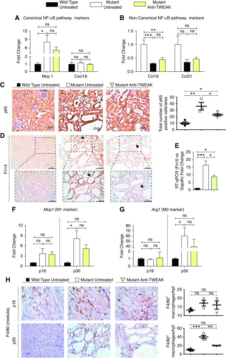
Anti-TWEAK treatment reduces NF-kB pathway activation and macrophage recruitment. Gene expression assessed by RT-qPCR of (A) Mcp1 and Cxcl16, and (B) Ccl19 and Ccl21, markers of canonic and noncanonic NF-κB pathway activation, respectively. Anti-TWEAK significantly decreases canonic NF-κB pathway activation. (C) Nuclear p65 staining is decreased in anti-TWEAK–treated mice. Representative images are shown. Mice euthanized at p30 (Supplemental Figure 4C). Anti-TWEAK decreases Fn14 expression in ADPKD. (D) Fn14 staining is decreased in cyst-lining epithelium (arrows) of mutant anti-TWEAK–treated mice. Mice euthanized at p30 (Supplemental Figure 4C). (E) Fn14 gene expression was diminished in anti-TWEAK–treated group. Mice euthanized at p30 (Supplemental Figure 4C). RT-qPCR of (F) Mcp1 (M1 markers) and (G) Arg1 (M2 markers) at p18 and p30. No differences were found at p18 (precystic stage), but anti-TWEAK treatment resulted in reduced gene expression of both proinflammatory markers at p30. Macrophage infiltration was reduced by anti-TWEAK treatment. (H) F4/80 staining in kidney tissue at p18 and p30. There were significantly reduced numbers of positive F4/80 macrophages in the medulla at p30. WT (n=6 at p18, n=5 at p30), mutant (n=6 at p18, n=5 at p30), and mutant mice treated with anti-TWEAK (n=7 at p18, n=6 at p30) were used for this RT-qPCR experiment set. Hprt was used as the housekeeping gene in all cases. For immunohistochemistry analysi s, n=4 was used per group in all instances. Bars represent means±SEM. *P<0.05, **P<0.01, ***P<0.001.

