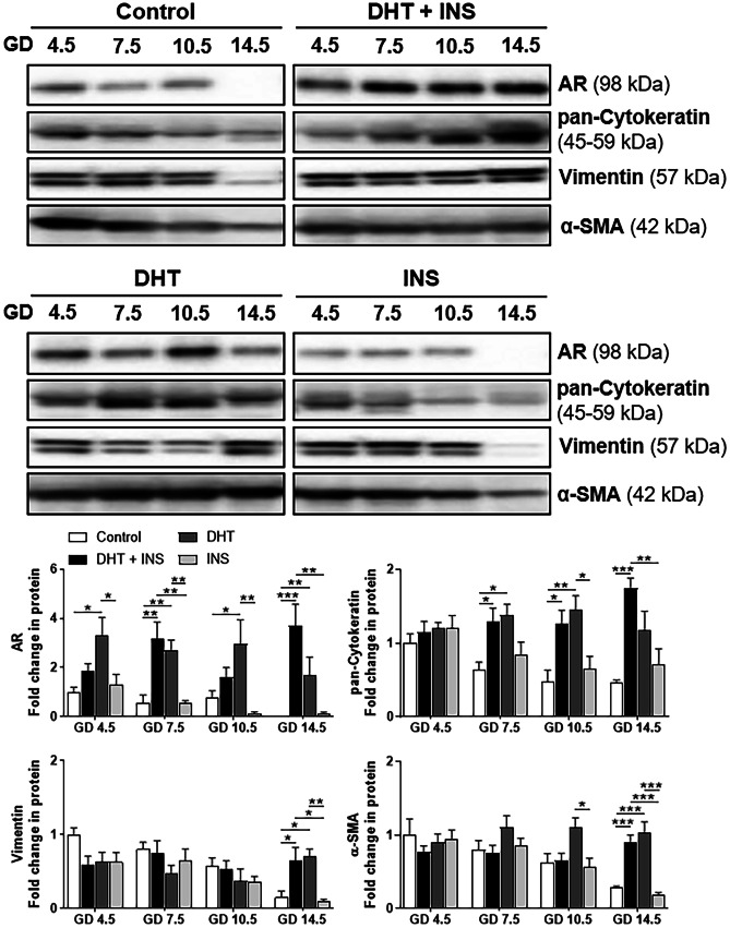
Fig. 2.
Differential regulation of AR protein in uteri collected from pregnant rats exposed to DHT and/or INS from GD 4.5 to GD 14.5. Time-dependent regulation of AR protein abundance in the pregnant uterus. After removing the embryos/fetuses and placenta, the uterine tissues from pregnant rats treated with control (vehicle), DHT + INS, DHT, or INS were used for analyzing AR and uterine cell marker proteins (cytokeratin, vimentin, and α-smooth muscle actin) by Western blotting. In all plots, the relative mean protein abundance ± SEM (vs. Control GD 4.5 values, n = 5–6/group) was measured by Western blotting with total proteins serving as loading controls. Statistical tests are described in the “Materials and methods” section, and differences between the groups are reported as *P < 0.05, **P < 0.01, and ***P < 0.001. The size representation in kilodaltons (kDa), as determined by a molecular weight ladder, is shown to the right

