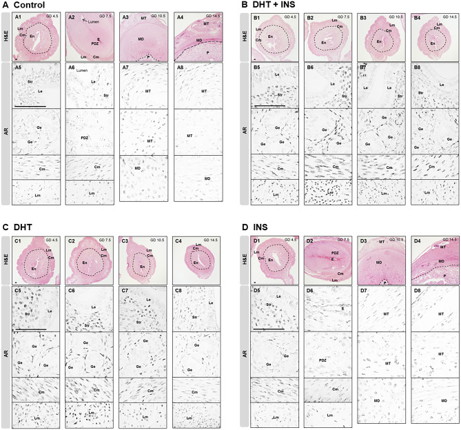
Fig. 3.
Localization of the AR protein in uteri collected from pregnant rats exposed to DHT and/or INS from GD 4.5 to GD 14.5. Histological appearance of gravid uteri using hematoxylin and eosin (H&E) staining (A1–A4, B1–B4, C1–C4, and D1–D4) and AR localization by immunohistochemistry (A5–A8, B5–B8, C5–C8, and D5–D8) in pregnant rats treated with vehicle A, DHT + INS B, DHT C, or INS D. Images are representative of eight tissue replicates. GD, gestational day; DHT, 5α-dihydrotestosterone; INS, insulin; En, endometrium; Cm, circular myometrium; Lm, longitudinal myometrium; Le, luminal epithelial cells; Ge, glandular epithelial cells; Str, stromal cells; E, embryo; PDZ, primary decidual zone; MT, mesometrial triangle; MD, mesometrial decidua; P, placental disc. Scale bars (100 μm) are indicated in the photomicrographs

