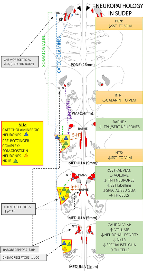
Figure 2. Medullary respiratory regulatory pathway and evidence for involvement in SUDEP.
The autonomic nuclei under study (as detailed in the text) have been highlighted only for simplicity (the autonomic and respiratory nuclei are shown in red and the ventrolateral medulla (VLM) region in yellow) and neuronal groups in VLM depicted as triangles; blue for catecholaminergic, orange for somatostatin neurones and green for NK1R positive neurones (neurokinin 1 receptor). The dashed lines indicate some of the known modulatory pathways shown on the left hand side. On the right hand side a summary of the cellular findings in SUDEP is detailed; green boxes are observations (see main text for detail) and orange boxes are changes inferred or hypothesised from observations but requiring substantiation in further studies. TH= tyrosine hydroxylase, SST= somatostatin, PBN= parabrachial nucleus, NTS= nucleus of tracus solitarius, RTN= retrotrapezoid nucleus

