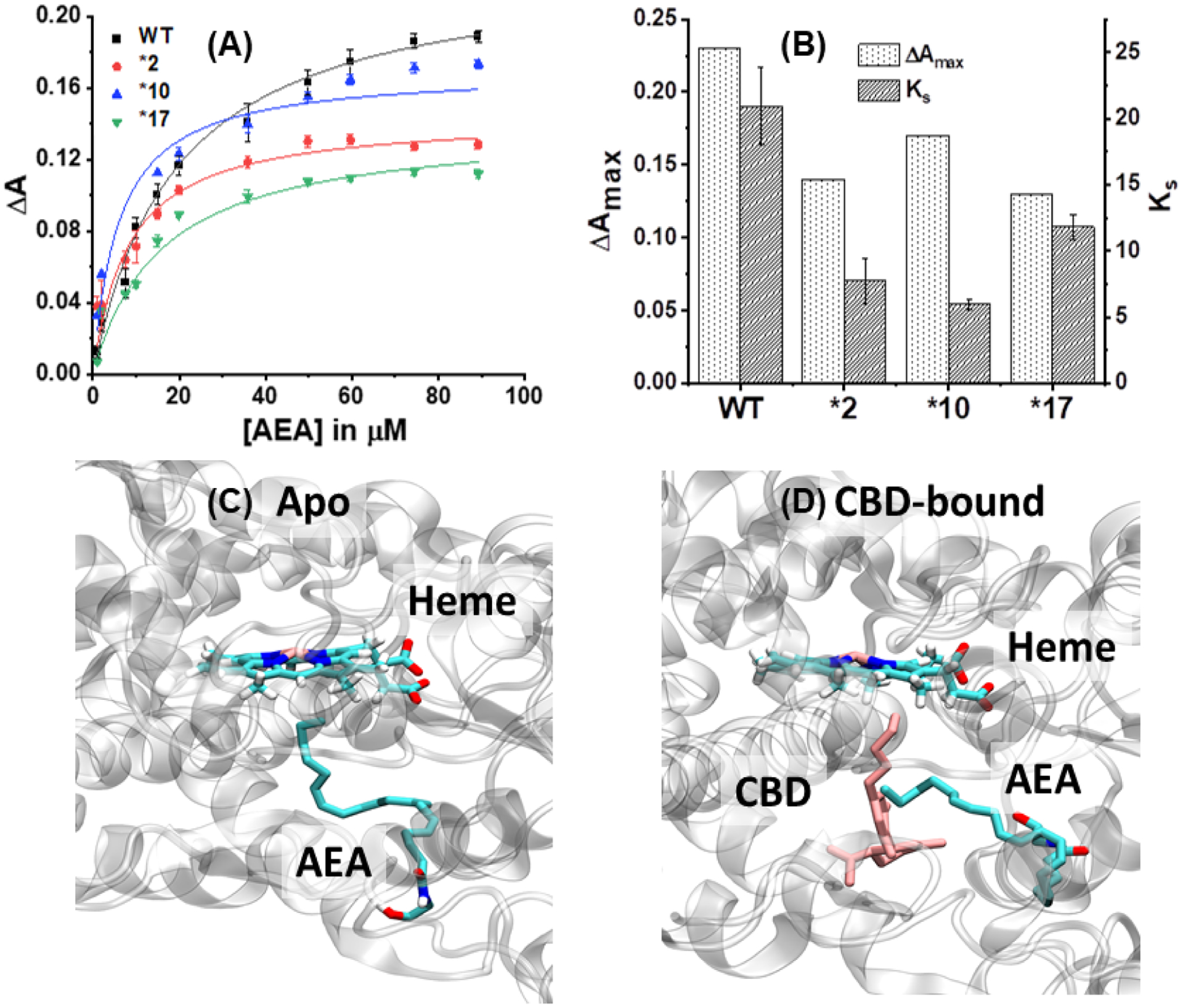
Figure 6:

Soret titration binding curves for (A) AEA in presence of WT, *2, *10 and *17. ΔA indicates the difference in absorbance between 393 nm and 417 nm. (B) Comparison of spin state change (ΔAmax) and Ks for AEA in presence of different 2D6 constructs. Docking studies showing the binding site for anandamide (AEA) in (C) Apo: AEA bound to CYP2D6; (D) CBD-bound WT CYP2D6. The heme, AEA and CBD are highlighted.
