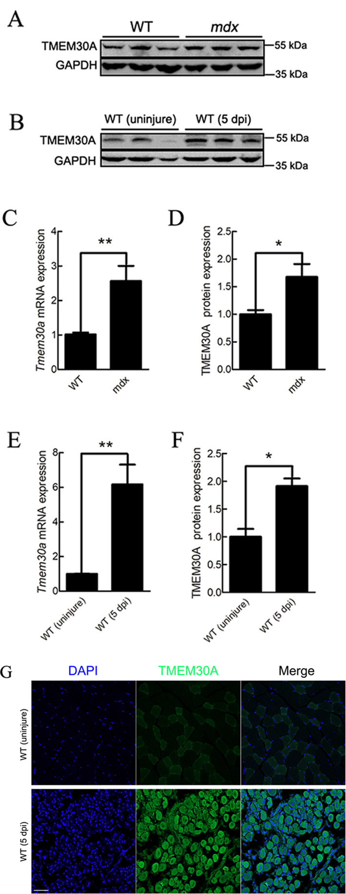
Figure 1.

Tmem30a was up-regulated during skeletal muscle regeneration
A, D: Immunoblot analysis of TMEM30A protein expression in 3-month-old wild-type (WT) and mdx mice. Relative protein density was calculated by ImageJ. B, F: Immunoblot analysis of TMEM30A protein expression in uninjured WT mice and in WT mice at 5 days post-injury (dpi). Relative protein density was calculated by ImageJ. C: mRNA expression analysis of Tmem30a in 3-month-old WT and mdx mice using qRT-PCR. E: mRNA expression analysis of Tmem30a in uninjured WT mice and in WT mice at 3 dpi using qRT-PCR. Data are mean±SEM. n=3 in each group. Significance was calculated using two-tailed Student’s t-test. *: P<0.05;**: P<0.01. G: Immunofluorescence analysis of TMEM30A expression in TA muscles of uninjured WT mice and of WT mice at 5 dpi. TMEM30A immunostaining is shown in green, and nuclei are shown in blue (counterstained with DAPI). Scale bar: 50 μm.
