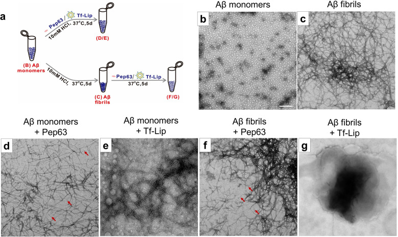
Fig. 4. Tf-Lip and Pep63 block the aggregation of Aβ1-42 monomers or disaggregate Aβ1-42 fibrils in vitro.
a Schematic diagram of the preparation of Aβ aggregates. b Electron micrograph of 100 μM Aβ1-42 monomers. c Aβ1-42 fibrils of 100 μM Aβ1-42 monomers formed after 5 days incubation with 10 mM HCl at 37 °C. d, e Final Aβ1-42 assemblies of 100 μM Aβ1-42 monomers formed after 5 days incubation with 2.5 μg/ml Pep63 (d) or 72.5 μg lipids/ml Tf-Lip (e) at 37 °C for 5 days. f, g Final Aβ1-42 assemblies of 100 μM Aβ fibrils obtained after incubation with Pep63 (f) or Tf-Lip (g) at 37 °C for 5 days. Arrows are indications for Pep63. Scale bar: 200 nm.

