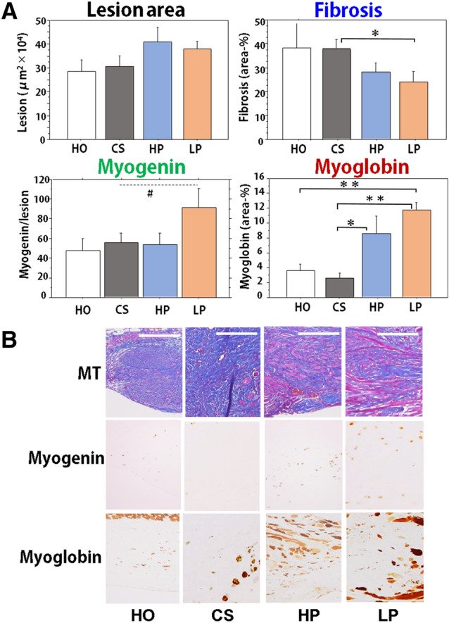
Figure 2.

Morphological alterations of granulation tissues after wounding of the abdominal wall in mice that consumed different peptides. (A) After the study was completed (day 21), the wound lesion on the abdominal muscle wall was examined for granulation area, fibrosis area, myoglobin positive regenerative muscle area, and myogenin positive cell number in the following groups, HO, CS, HP, and LP. N = 5 or 6/group, Mean ± SE, #p > 0.1, *p < 0.05, **p < 0.01. (B) Representative lesions from the HO, CS, LP, and HP groups (day 21). Serial sections were stained by MT stain and immunobiological stains for myogenin and myoglobin. Bars = 100 µm.
