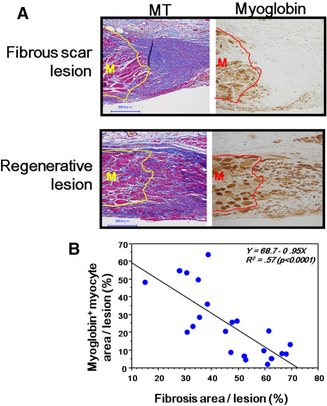
Figure 5.

The relation of the fibrous scar lesion and muscle regeneration in wound healing in the abdominal muscle wall. (A) The representative lesions of the fibrous scarring and muscle regeneration stained by MT and myoglobin staining using serial sections; The lesion in the upper panel was from the control group on day 21, and the lesion in the lower panel was from the Pro-Hyp group on day 21. The lower panel shows the stumped muscle (M), from which regenerated muscle cells migrate into the granulation tissue. Bars = 200 µm. (B) The relation of fibrosis area (%) and myoglobin positive area (%) in the granulation tissue obtained from both groups on day 21.
