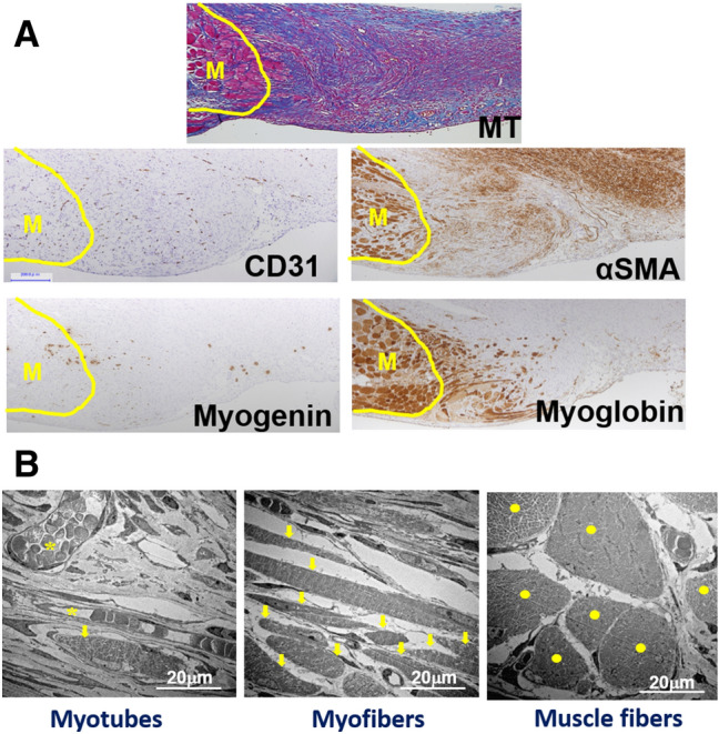
Figure 6.

Muscle regeneration from the stumped muscle wall in granulation tissue. (A) Distribution of CD31, αSMA, myogenin, and myoglobin positive cells are shown in the granulation tissue with muscle regeneration. The seral sections were obtained from the Pro-Hyp group on day 14. Bars = 200 µm. (B) Electron micrographs of the abdominal lesion obtained from the Pro-Hyp group on day 14 showing myogenic differentiation in the granulation tissue. Myotubes forming a multinuclear envelope (left, arrows). Elongated myofibers with transverse striations (meddle, arrows). Large matured muscle fibers (right, dots). Bars = 20 µm.
