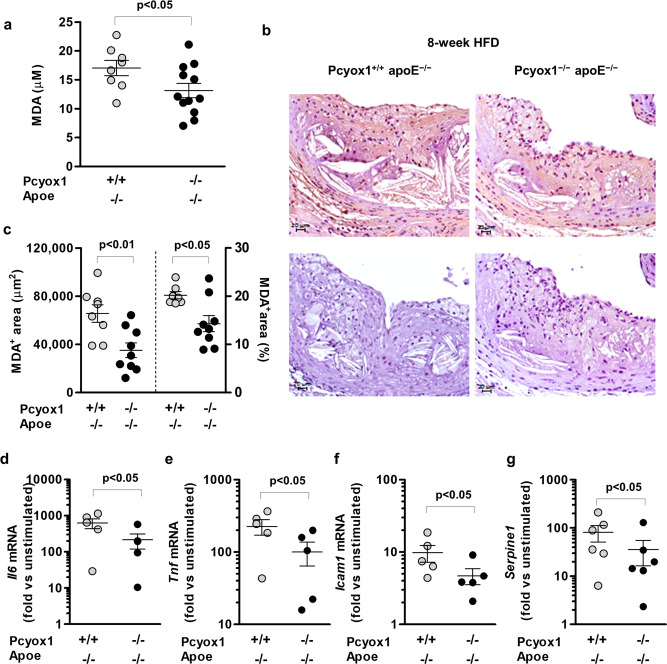
Fig. 6. Pcyox1 deficiency is associated with lower levels of lipid peroxidation and decreased LPS-induced inflammatory response in murine peritoneal macrophages.
MDA in plasma (a) and in the aortic root lesions (b, c) from Pcyox1+/+/Apoe−/− (n = 8) and Pcyox1−/−/Apoe−/− (n = 11) mice fed a HFD for 8 weeks. b Representative images of MDA immunostaining (upper panels) and negative control sections incubated with no primary antibody (lower panels); c results of the morphometric analysis of MDA-stained sections, expressed either as absolute positive areas within the intima or as the percentage of total lesion area. Data are presented as circle plot, with each circle representing an individual mouse and bars showing the mean value ± SEM. p < 0.05 or p < 0.01 by Student’s t test. d–g mRNA levels normalized to the housekeeping gene 18S rRNA of Il6 (d), Tnf (e), Icam1 (f), and Serpine1 (g) in macrophages isolated from Pcyox1+/+/Apoe−/− and Pcyox1−/−/Apoe−/− mice and treated with LPS 5 ng/mL for 2 h. Data are expressed as fold increase induced by LPS with respect to unstimulated cells and presented as circle plot, with each circle representing an individual mouse and bars showing the mean value ± SEM (n = 5). p < 0.05 by Student’s t test.

