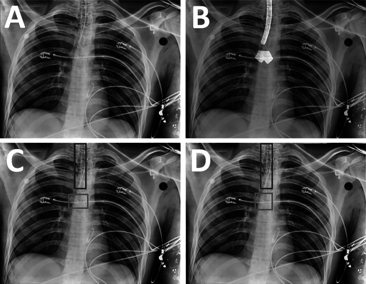
Fig. 2.
Generation of labeled training data from a CXR. A The original image. B Segmentations of the endotracheal tube and carina are drawn on the image by radiologists using a brush tool (white overlays). C The outer perimeters of the segmentations are used to obtain bounding boxes for the endotracheal tube (blue box) and carina (red box). D The carina box (red box) is modified to make it a square of fixed size in proportion to the image size

