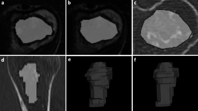
Fig. 1.
Contour-focused and margin shrinkage segmentation of an atypical cartilaginous tumor of the humerus in a 45-year-old woman. a–c 2D contour-focused segmentation was performed on axial T1-weighted MRI a, T2-weighted MRI b, and bone-window CT c on the slice showing the largest tumor extension. d 3D contour-focused segmentation was performed slice by slice in the axial plane to include the whole tumor volume, as shown in the sagittal CT image. Contour-focused segmentation provided the ROI including both green and red areas. Margin shrinkage segmentation provided the ROI including only the green area by computing a marginal erosion, which is shown in red. e–f Segmented tumor volumes obtained with 3D contour-focused e and margin shrinkage f segmentation are shown, where the latter has smoother margins as a result of marginal erosion

