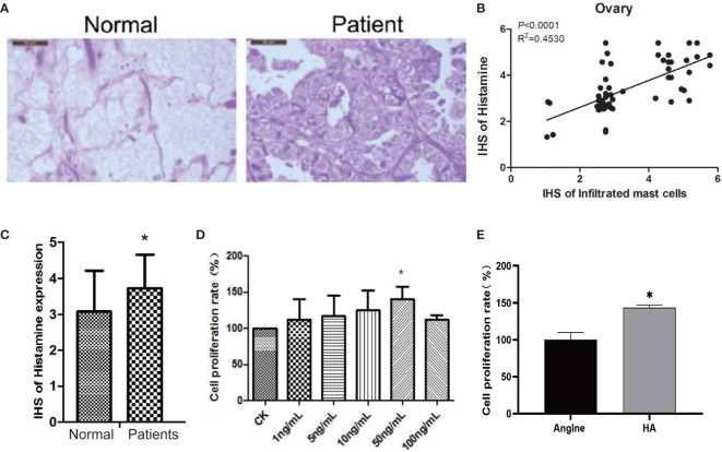
Figure 2.
Histamine released by mast cell degranulation stimulates the proliferation of ovarian cancer cells. (A) HE staining of normal ovarian tissue (left) and ovarian cancer tissue (right). (B) Correlation between mast cell infiltration and histamine release in ovarian cancer tissue. (C) Histamine release levels in normal ovarian and ovarian cancer tissues, *P < 0.05 (compared to normal tissues). (D) OVCAR-3 cell proliferation after treatment with different concentrations of histamine. (E) Anglne cell proliferation rate after treatment with histamine at a concentration of 50 ng/mL. *P < 0.05 (compared to untreated Anglne cell group).

