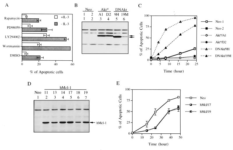
FIG. 1.
The PI3-K/Akt kinase pathway is involved in the viability response of IL-3 in Ba/F3 cells. (A) Ba/F3 cells cultivated in growth medium with or without IL-3 were treated with various chemical inhibitors as indicated. Fifteen hours after each treatment, cells were harvested and the percentage of apoptotic cells under each condition was quantified by flow cytometric analysis of cells with a sub-G1 DNA content. Results shown are means ± standard deviations from three independent experiments. (B) Protein expression of Ba/F3 cells overexpressing M-Akt (Ba/F3Akt*) or AktK179M (Ba/F3DNAkt). One hundred micrograms of protein lysates from Ba/F3Akt* (clones A1 and D2) or from Ba/F3DNAkt (clones 9H and 19M) were analyzed by immunoblotting with anti-HA antibody (both proteins are HA tagged). Specific bands are indicated by arrows. (C) Ba/F3 derivatives as indicated were deprived of IL-3, and the percentage of apoptotic cells at various time points was measured as described in panel A. During IL-3 deprivation, these cells were kept in medium containing 10% instead of 0.5% FBS (see Materials and Methods) to slow the death rate of Ba/F3DNAkt cells. Results shown here are the averages of two independent experiments. (D) Same as in panel B except that the cells analyzed were Ba/F3 cells stably overexpressing human Mcl-1 protein (Ba/F3hMcl-1) and the immunoblot probed with anti-human Mcl-1 antibody, which did not cross-react with the murine homologue. Several individual clones, as indicated by numbers, were analyzed. (E) Ba/F3hMcl-1 cells were deprived of IL-3 by the standard procedure as described in Materials and Methods, and the percentage of cells undergoing apoptosis at various time points was analyzed as described in panel A. Only results from two representative clones (hMcl/17 and hMcl/19) are shown. Neo represents control cells that are stably transfected by the empty expression vector and is a mixture of two stable lines. Results shown are means ± standard deviations from two independent experiments.

