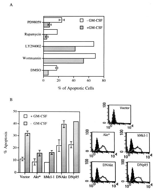
FIG. 2.
The PI3-K/Akt kinase pathway is involved in the viability response of GM-CSF in TF-1 cells. (A) TF-1 cells cultivated in growth medium with or without GM-CSF were treated with various inhibitors for 15 h and analyzed as described in the legend to Fig. 1A. The percentage of apoptotic cells under each condition was expressed as the mean ± standard deviation from two independent experiments. (B) TF-1 cells were transiently transfected with the constructs of interest plus GFP expression vectors. Twenty-four hours after transfection, cells were placed in medium with or without GM-CSF for another 18 h before they were fixed, stained, and analyzed as described in Materials and Methods. The GFP-positive and annexin-V bound (apoptotic) cells were quantified by flow cytometry, and the results are plotted in the left panel. The right-hand panel displays the flow cytometric results showing expression of each individual protein in the transfected (GFP positive, gray lines), but not in the nontransfected (GFP negative, solid peaks), fractions. All proteins were HA tagged and detected by HA-specific antibody. Vector, Akt*, hMcl-1, DNAkt, and DNp85 denote cells transfected with an empty expression vector or vectors expressing M-Akt, human Mcl-1 protein, AktK179M, and the dominant negative mutant of PI3-K (Δp85), respectively.

