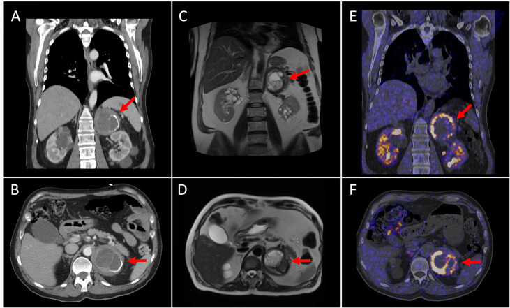
Figure 2.
Coronal and axial CT views (A, B) show an 8 cm left adrenal tumor with a cystic component inside and partially calcified wall. Coronal and axial MRI views (C, D) reveal a left adrenal mass with a heterogeneous content (necrotic-cystic areas). Coronal and axial [18 F] DOPA PET/CT images (E, F) demonstrate intense and heterogeneous DOPA-uptake in the periphery of the mass (SUVmax of 20.9). These findings were compatible with the existence of pheochromocytoma with a cystic–necrotic–hemorrhagic component.

