Figure 4.

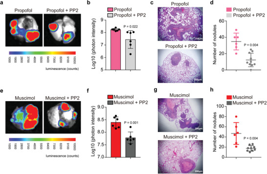
Inhibition of Src attenuates the propofol‐ or GABAAR agonist‐promoted tumor metastasis in mice lungs. a) Ex vivo bioluminescent assay of lung tumor metastasis. HCT116 cells overexpressing luciferase were treated with propofol or propofol plus PP2 (inhibitor of Src) for 3 h. Treated cells were then injected intravenously into BALB/c nude mice through the tail vein. b) Quantification of bioluminescent photon intensity (N = 6 in each group; mean ± SD, Student's t‐test, P = 0.022). c) H&E staining of metastatic lung nodules (scale bar = 200 µm). d) Quantification of metastatic nodules (N = 6 in the propofol group, N = 10 in the propofol plus PP2 group; median and IQR, Mann–Whitney test, P = 0.004). e) Ex vivo bioluminescent assay of lung tumor metastasis. HCT116 cells overexpressing luciferase were treated with muscimol or muscimol plus PP2 for 3 h. Treated cells were then injected intravenously into BALB/c nude mice. f) Quantification of bioluminescent photon intensity (N = 6 in each group; mean ± SD, Student's t‐test, P = 0.001). g) H&E staining of metastatic lung nodules (scale bar = 200 µm). h) Quantification of metastatic nodules (N = 6 in the muscimol group, N = 9 in the muscimol plus PP2 group; median and IQR, Mann–Whitney test, P = 0.004). VEC, vein endothelial cell; H&E, hematoxylin and eosin; SD, standard deviation.
