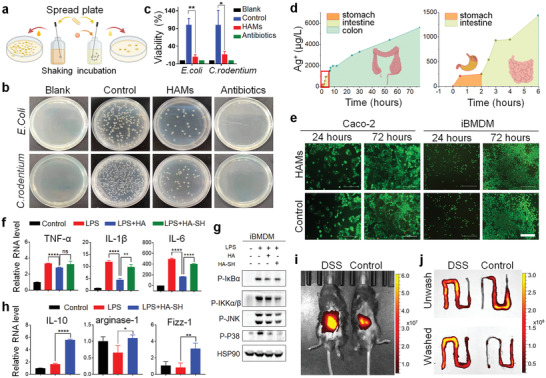
Figure 2.

Characteristics of HAMs. a) Schematic illustration of the anti‐bacterial experiment. b) The anti‐bacterial activity of HAMs was shown by the spread plate method. c) The bacterial viability of Escherichia coli (E. coli) and Citrobacter rodentium (C. rodentium) in (b) were measured by colony count. The experiment was repeated three times. d) The silver ion (Ag+) release from 100 mg HAMs in 1 mL artificial gastric, intestinal, and colon fluids was detected by inductively coupled plasma mass spectrometer. e) The cytotoxicity of HAMs was examined in Caco‐2 and immortalized bone marrow‐derived macrophage (iBMDM) by using the calcein acetoxymethyl ester/propidium iodide cell viability kit. Scale bar 500 µm. f) mRNA expression of the cytokines including interleukin‐6 (IL‐6), interleukin‐1β (IL‐1β), and tumor necrosis factor‐α (TNF‐α). g) Western blot analysis of protein level of phosphorylated inhibitor of nuclear factor κB (P‐IκBα) and phosphorylated inhibitory kappa B kinase α/β (P‐IKKα/β), phosphorylated‐P38 and phosphorylated c‐Jun N‐terminal kinase (P‐JNK) in iBMDM with different treatment. h) mRNA levels of M2 macrophage‐related genes, including interleukin‐10 (IL‐10), arginase‐1, and found in inflammatory zone‐1 (Fizz‐1). i,j) Healthy mice and mice with dextran sulfate sodium (DSS)‐induced colitis were imaged by in vivo imaging system after the oral gavage of indocyanine green (ICG)‐HAMs, and the intestine with strong signals was removed and then imaged before and after washing. The experiments were repeated three times. Significance between every two groups was assessed by using Mann–Whitney U‐test. ns, not significant; * p < 0.05, ** p < 0.01, **** p < 0.0001.
