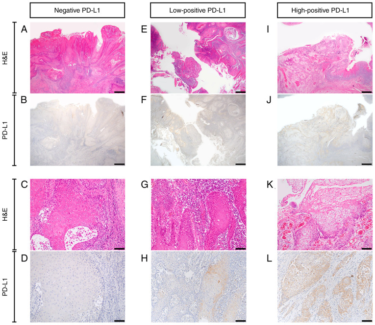
Figure 3.
PD-L1 expression in oral squamous cell carcinoma. (A-D) Negative PD-L1 expression. (E-H) Low-positive PD-L1 expression. (I-L) High-positive PD-L1 expression. (A, C, E, G, I and K) H&E staining. (B, D, F, H, J and L) Immunohistochemistry staining for PD-L1. Scale bar=1 mm in A, B, E, F, I and J. Scale bar=100 µm in C, D, G, H, K and L. PD-L1, programmed death ligand-1; H&E, hematoxylin and eosin.

