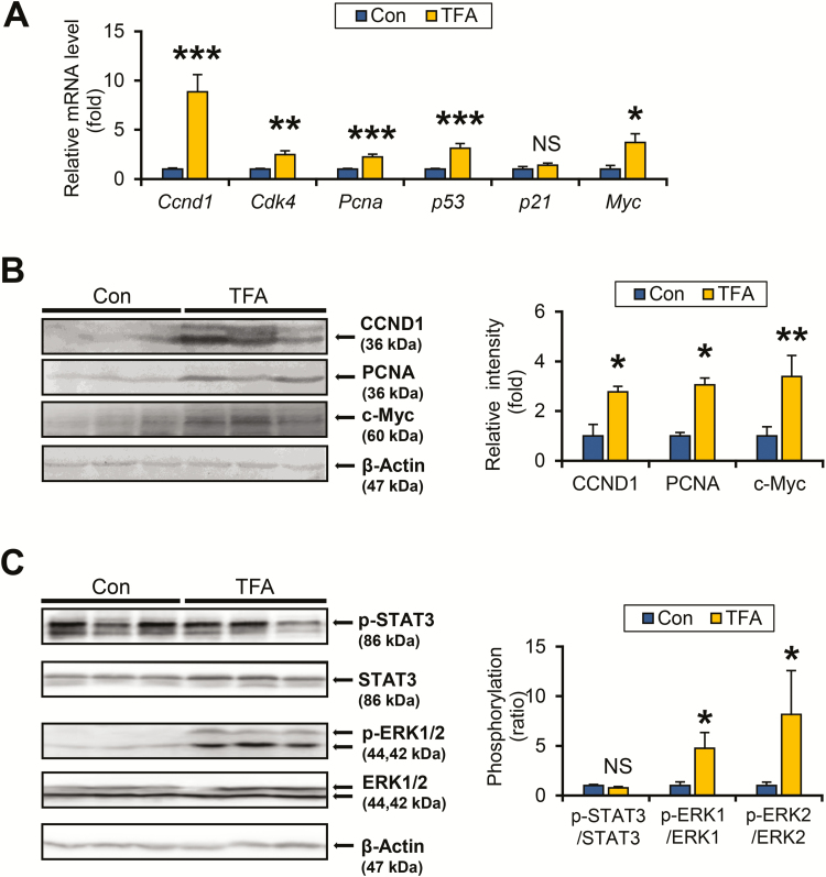
Figure 5.
TFA-rich diet promotes ERK phosphorylation and hepatocyte proliferation in HCVcpTg mice. (A) Hepatic mRNA levels of cell cycle regulators were quantified by qPCR, normalized to those of 18S ribosomal RNA and expressed as values relative to HCVcpTg mice fed a control diet. (B) Immunoblot analysis of CCND1, proliferating cell nuclear antigen and c-Myc. Whole-liver homogenates (60–80 μg of protein) were loaded into each well and the band of β-actin was used as a loading control. Band intensities were measured densitometrically, normalized to those of β-actin and expressed as values relative to HCVcpTg mice fed a control diet. Results were obtained from two independent immunoblot experiments and representative blots were shown. (C) Immunoblot analysis of STAT3 and ERK1/2. Whole-liver homogenates (45 μg of protein) were loaded into each well and the band of β-actin was used as a loading control. Band intensities were measured densitometrically, normalized to those of β-actin, calculated as phosphorylated/total ratio values and expressed as values relative to HCVcpTg mice fed a control diet. Results were obtained from two independent immunoblot experiments and representative blots were shown. Data are expressed as mean ± SEM. *P < 0.05, **P < 0.01 and ***P < 0.001 between control diet-fed and TFA-rich diet-fed HCVcpTg mice. Con, control diet-fed HCVcpTg mice; TFA, TFA-rich diet-fed HCVcpTg mice; NS, not significant; p-, phosphorylated.

