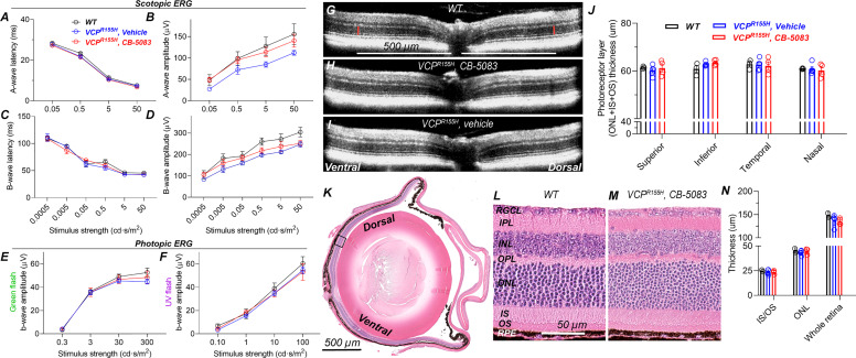
Fig. 4.
Chronic use of CB-5083 is safe for the eyes, and visual function recovers after drug washout. VCP-disease mice (VCPR155H knock-in heterozygous mutants) were treated daily with CB-5083 (n = 4, 15 mg/kg) or with vehicle (controls, n = 5) by gavage for 6 months between 12 and 18 months of age. Eighteen-month-old, healthy, and nontreated C57BL/6J mice were used as a WT control (n = 3). At the end of the trial, ERG and OCT were conducted 24 hours after the last dose. (A and C) Scotopic ERG A- and B-wave latency analyses indicate no difference in phototransduction or photoreceptor-to-bipolar cell synaptic transmission speed, respectively, between the study groups. Instead, scotopic ERG amplitude analyses (B and D) indicate moderately but statistically significantly lower A- and B-wave amplitudes for vehicle-treated VCP mice compared with nontreated healthy controls (2-way ANOVA between-subjects main effects in both: P < 0.05). CB-5083–treated VCP mice had slightly higher B-wave amplitudes compared with vehicle-treated VCP mice (ANOVA between-subjects: P < 0.05). (E and F) Photopic ERG amplitudes did not differ between study groups. (G–I) Representative OCT images from WT (G), CB-5083–treated (H), and vehicle-treated (I) VCP-disease mice. (J) Photoreceptor thickness is equal among the three study groups. (K) Representative histologic WT mouse eye section for illustration. The black box indicates the area where 40× images (L and M) were acquired. (L and M) High-magnification images show similar morphology in the nontreated WT and chronically CB-5083–treated VCP-disease mouse retinas. (N) IS/OS, ONL, and whole retina thickness measurements from histology images (n for CB-5083–treated same as above, but n = 2 for WT). INL, inner nuclear layer; IPL, inner plexiform layer; OPL, outer plexiform layer; RGCL, retinal ganglion cell layer; RPE, retinal pigment epithelium. All graphs show mean ± S.D., and retinal thickness layer analyses also show individual replicates. Statistical comparison was performed using 2-way (ERG and OCT) or 1-way ANOVA (histology).

