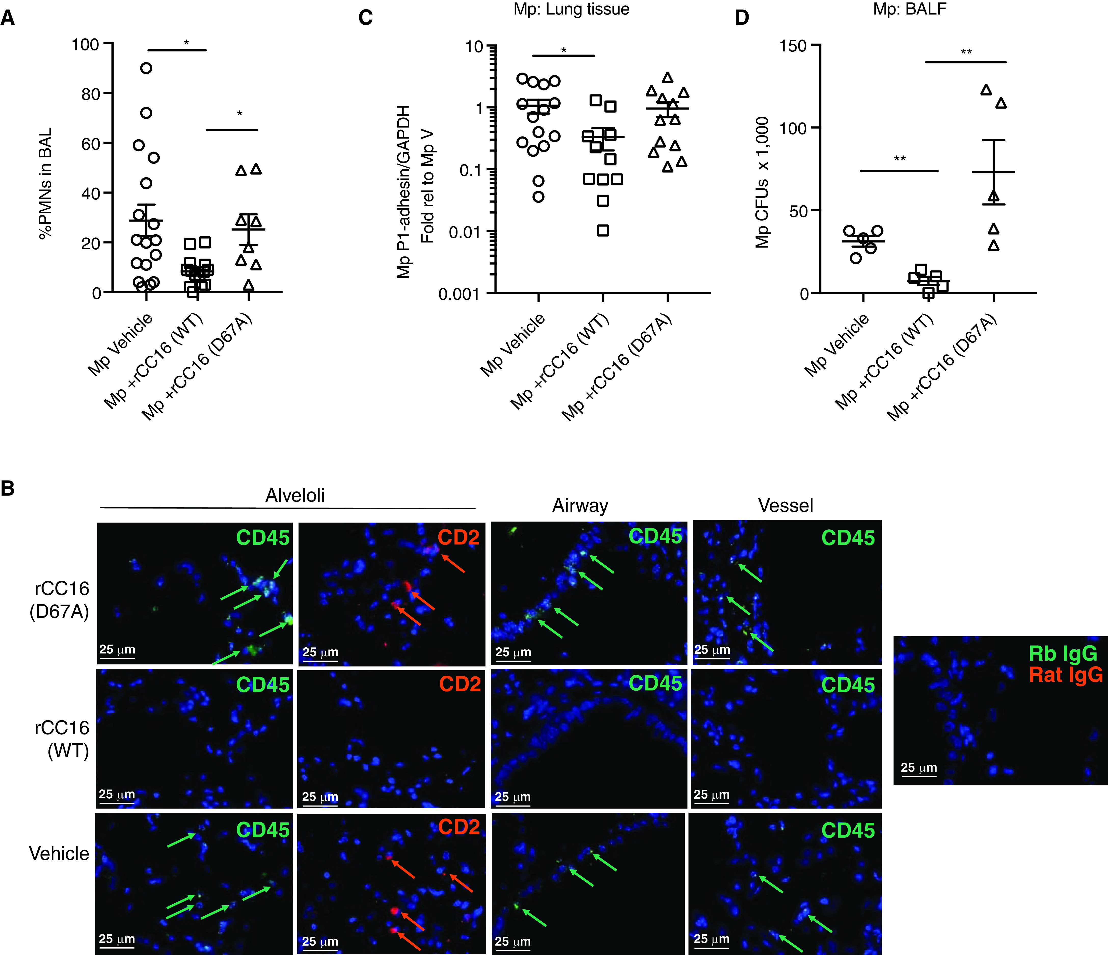
Figure 4.

Inflammatory cell assessment and pathogen burden based on CC16 (club cell secretory protein) treatment. (A) A subset of Cc16−/− mice that were assessed for pulmonary function tests were also assessed for the presence of neutrophils in the lavage fluid by differential staining. CC16-deficient mice that received recombinant CC16 (rCC16) (WT) had a significantly lower percentage of neutrophils (PMNS) versus those that received no CC16 or rCC16 (D67A). (B) Representative (n = 4/treatment group) immunofluorescence staining for CD45 and CD2 in alveoli, airways, and vessels in the specific treatment groups. Arrows indicate positively stained cells relative to negative antibody controls (Rb IgG and rat IgG). Scale bars, 25 μm. (C) Assessment of Mycoplasma pneumoniae (Mp) burden in lung tissue by RT-PCR for Mp-specific P1-adhesin gene relative to GAPDH. Data shown as fold relative to Mp vehicle. (D) Assessment of Mp CFUs present in cell-free BAL at time of harvest. *P < 0.05 and **P < 0.01 by one-way ANOVA with Kruskal-Wallis test for multiple comparisons. BALF = BAL fluid; CFU = colony-forming unit; PMNS = polymorphonuclear leukocytes; V = vehicle; WT = wild-type.
