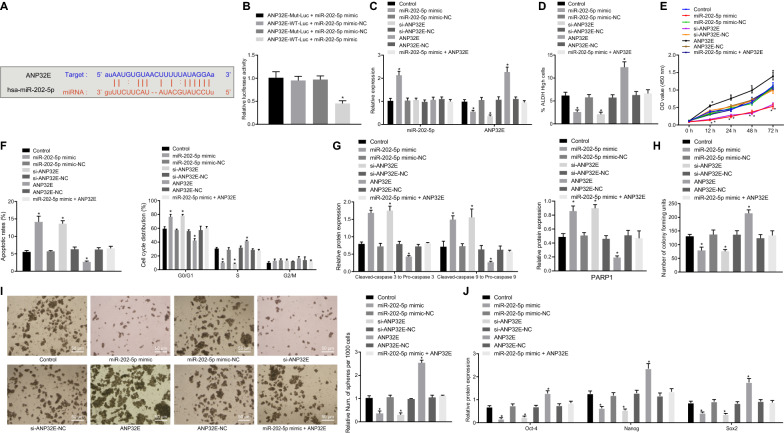
Fig. 3.
miR-202-5p inhibits the viability, proliferation, and self-renewal of PCSCs, and promotes cell apoptosis by suppressing ANP32E expression. A Potential target gene of miR-202-5p predicted by mirDIP, DIANA, TargetScan and starBase databases; B Targeting relationship between miR-202-5p and ANP32E measured by dual-luciferase reporter assay (* p < 0.05 vs. cells co-transfected with ANP32E-WT-Luc and miR-202-5p mimic-NC, ANP32E-MUT-Luc and miR-202-5p mimic, ANP32E-MUT-Luc and miR-202-5p mimic-NC). PCSCs were treated with ANP32E, si-ANP32E and/or miR-202-5p mimic. C miR-202-5p expression and the mRNA expression of ANP32E in PCSCs measured by RT-qPCR; D ALDH activity of PCSCs assessed by Aldefluor assay, where mock means a NC with the addition of DEAB (a specific inhibitor of ALDH enzyme); E Proliferation of PCSCs detected by MTT; F Apoptosis and cell cycle changes of PCSCs measured by flow cytometry; G Protein expression of PARP1 and the ratios of cleaved-caspase3 to pro-caspase3 and cleaved-caspase9 to pro-caspase9 in PCSCs detected by Western blot analysis; H Colony formation of PCSCs assessed by colony formation assay; I Self-renewal ability of PCSCs detected by sphere formation assay (200×); J Protein expression of Oct4, Nanog, Sox2 in PCSCs measured by Western blot analysis; * p < 0.05 vs. cells without treatment. Measurement data were expressed as mean ± standard derivation. Data among multiple groups were analyzed by one-way analysis of variance with Tukey’s post hoc test, and data comparison among multiple groups at different time points was conducted using two-way analysis of variance with Bonferroni post hoc test. The experiment was repeated three times

