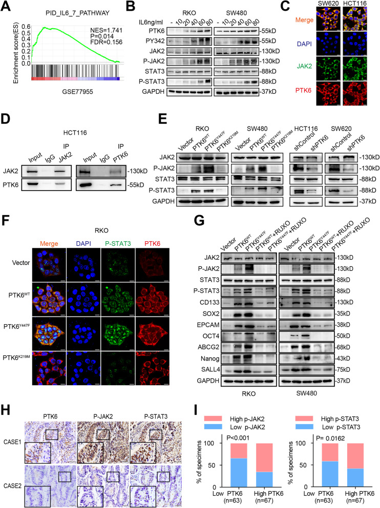
Fig. 5.
PTK6 promotes CRC progression by activating JAK2/STAT3 signaling pathway. A The GSEA result indicates an enrichment of gene sets related to IL6 signaling pathway in PTK6 overexpression group. B Western blot assays indicate that IL6 can stimulate the activation of the PTK6-JAK2/STAT3 pathway in a dose-dependent manner in CRC cells. C The co-localization of JAK2 (green) and PTK6 (red) in CRC cells was assessed by IF staining. The scale bar represents 5 μm. D Co-IP results show an endogenous protein interaction between PTK6 and JAK2 in CRC cells. E Western blot assays show that the phosphorylation of PTK6 plays a vital role in the activation of the JAK2/STAT3 pathway. F The co-localization of p-STAT3 (green) and PTK6 (red) in vector, PTK6WT, PTK6KM and PTK6YF overexpression CRC cells were assessed by IF staining. The scale bar represents 5 μm. G Western blot analyses show the expression of JAK2/STAT3 pathway members and stem cell markers in vector, PTK6WT, PTK6KM and PTK6YF overexpression CRC cells with or without the treatment of RUXO. H Representative immunohistochemical (IHC) staining images of CRC tissues from the Nanfang cohort show the expression of PTK6, p-JAK2 and p-STAT3 in CRC and the adjacent normal tissues. Scale bars represent 50 μm. I Histograms show that the expression of PTK6 is correlated with that of p-JAK2 and p-STAT3 in CRC tissues

