
Figure 6.
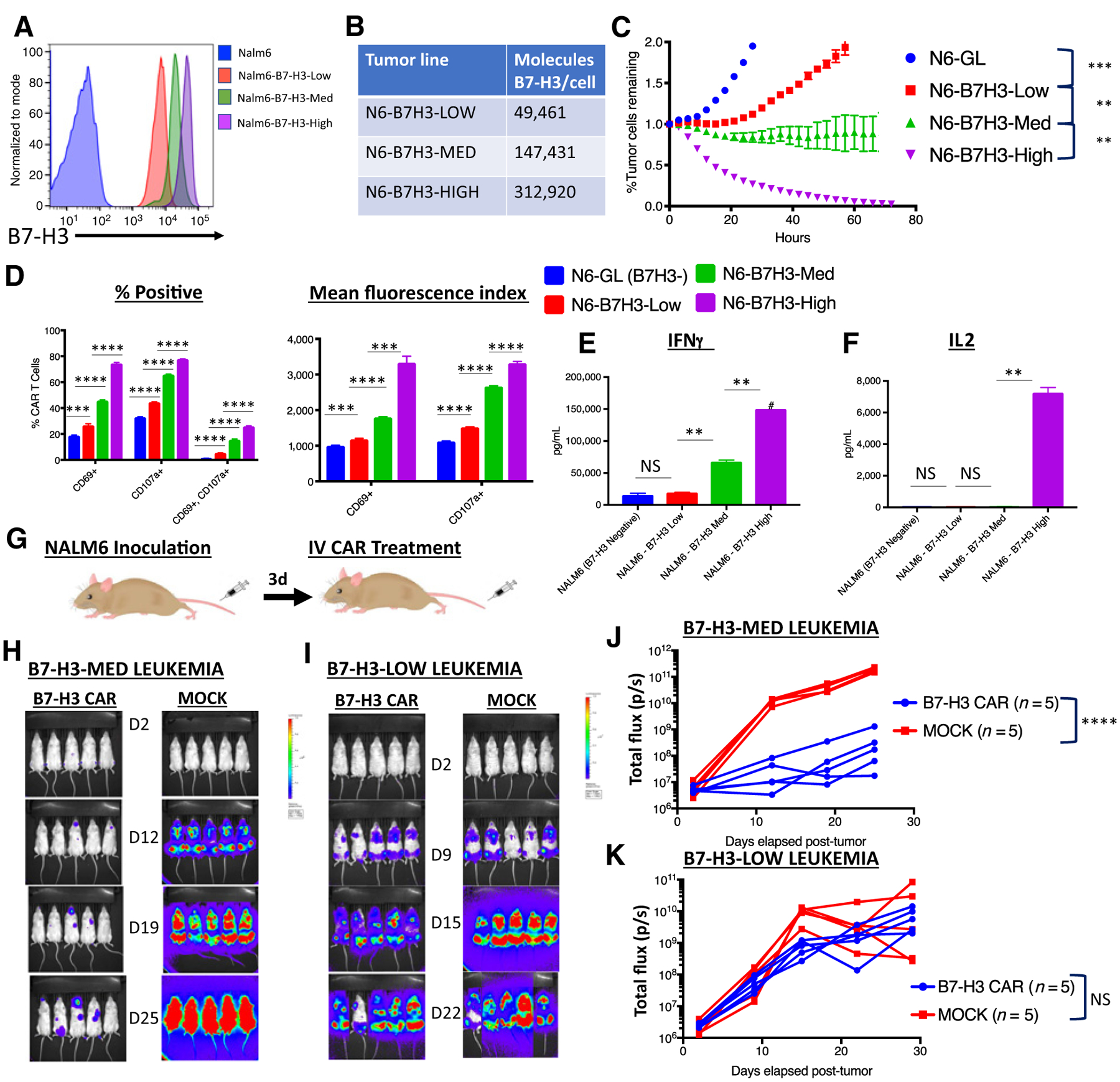
B7-H3 CAR T cells require adequate antigen expression for in vitro and in vivo activity. A, Flow cytometry analysis of B7-H3 expression on single-cell clones derived from Nalm6 expressing different amounts of lentivirally expressed B7-H3. B, Number of B7-H3 surface molecules expressed by Nalm6-B7-H3 cell lines as estimated by Quantibrite kit. C, GFP+ Nalm6-B7H3 clones were cocultured with B7-H3 CAR T cells and tumor cell killing was measured in an Incucyte assay over 72 hours. Representative data of three experiments with three different PBMC donors is shown. D, Percentage of CAR T cells positive (left) and mean fluorescence index (right) for T-cell activation and degranulation markers CD69 and CD107a, as measured by flow cytometry 6 hours after coculture of B7-H3 CAR T cells with tumor cells expressing increasing amounts of B7-H3. Representative results of three experiments with three different PBMC donors are shown. E and F, Cytokine production by CAR T cells cocultured with tumor cells expressing increasing amounts of B7-H3. G, Mouse model for Nalm6-B7H3: 1e6 NALM6 cells expressing either low or medium amounts of B7-H3 were engrafted into mice by tail vein injection. Three days later, mice were injected with 1e7 B7-H3 CAR+ T cells or untransduced control T cells (MOCK). In vivo imaging of mice bearing (H) Nalm6-B7-H3-Medium leukemia or (I) Nalm6-B7-H3-Low leukemia. J and K, Tumor progression was measured by bioluminescence photometry and flux values (photons per second) were calculated using Living Image software. Values for individual mice are shown. Representative results of four (Nalm6-B7-H3-Med) and two (Nalm6-B7-H3-Low) experiments with two different PBMC donors are shown. N6, NALM6; GL, GFP-Luciferase.