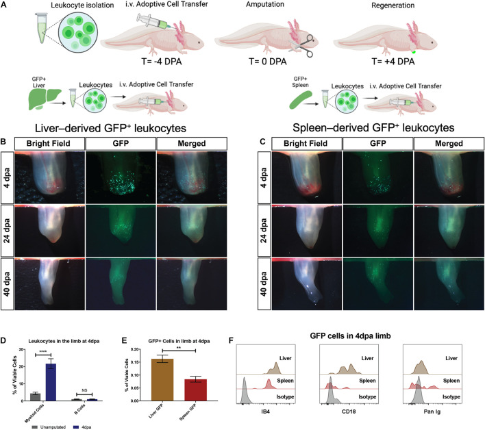
FIGURE 4.
The liver is the dominant contributor of myeloid cells during limb regeneration. (A) Experimental strategy to identify cellular contributions from the liver and spleen following transplantation of 5 × 10⁵ live GFP+ cells. (B,C) Representative live imaging of the regenerating host limb at the wound healing (4 dpa), blastema outgrowth (24 dpa), and re-development (40 dpa) stages of limb regeneration. Liver-derived GFP+ cells are most abundant during the wound healing stage (4 dpa) and reduce in number throughout later stages of regeneration. Spleen derived GFP cells are recruited to the limb but qualitatively less in number compared to liver transplanted cells. N = 12 biological replicates. (D) Quantitation of myeloid cells and B cells in the limb at 4 dpa using CD18/IB4 and Pan-Ig staining, respectively. Bar graphs show mean ± SEM of four biological samples. p-Values obtained via one-way ANOVA with comparisons to the unamputated sample. ****p ≤ 0.0001. (E) Quantitation of viable GFP+ cells in the early regenerating limb following liver or spleen transplantation. Bar graphs show mean ± SEM of three biological samples. p-Values obtained via one-way ANOVA. **p ≤ 0.01. (F) Flow cytometry histograms of GFP+ cells in the limb testing for myeloid cell identity (CD18) in the second plot and B cell identity in third plot. T, time; DPA, days post amputation. ns, not significant.

