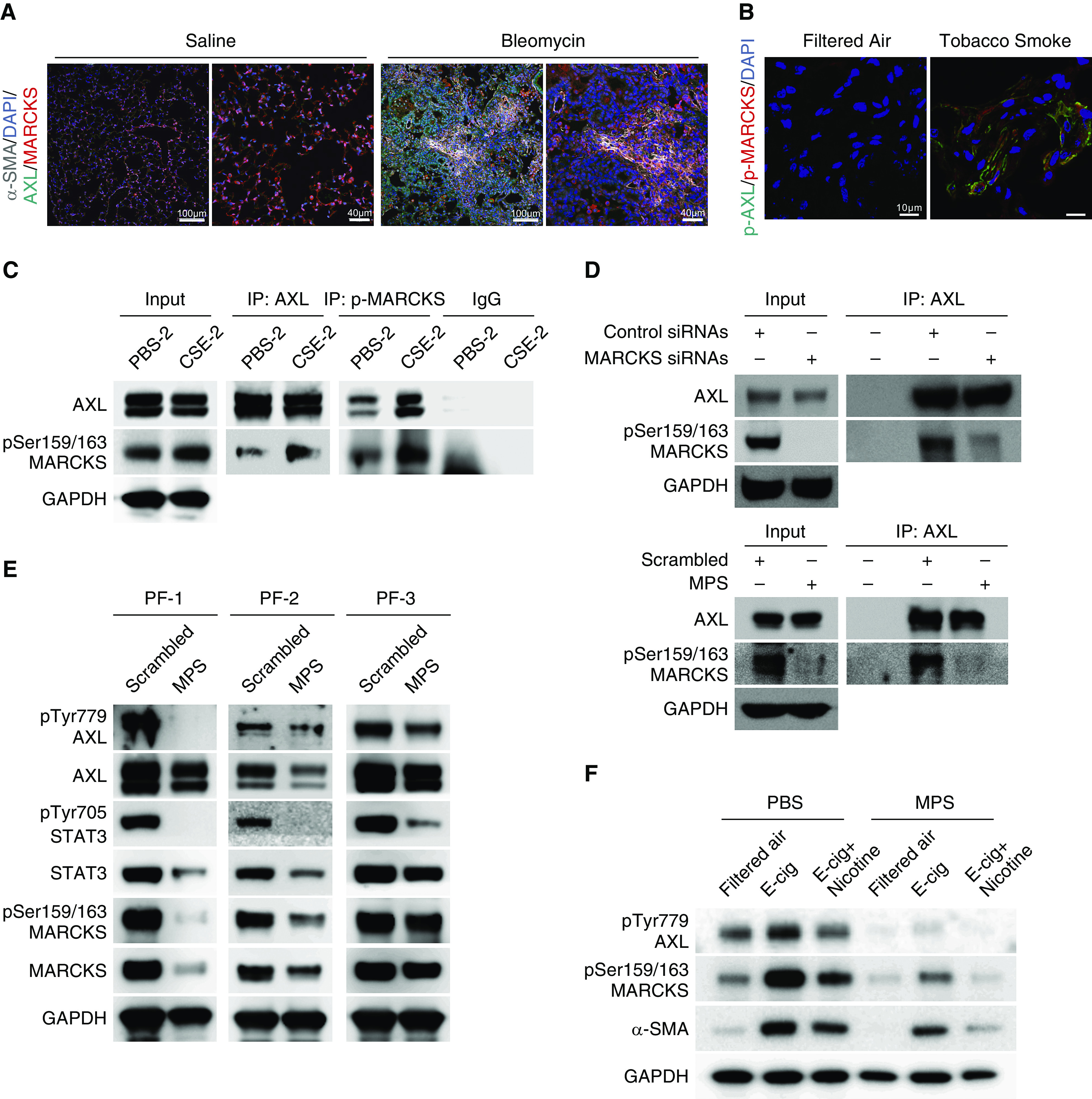
Figure 5.

AXL is associated with MARCKS in activated lung fibroblasts. (A) Representative immunofluorescence images of α-SMA (gray color), AXL (green color), and MARCKS (red color) in lung tissues from mice exposed to saline or bleomycin. DAPI (blue color): nuclear stains. Scale bars, 40 μm and 100 μm. (B) Representative immunofluorescence images of phospho-AXL (green color) and phospho-MARCKS (red color) in lung tissues from mice with exposure to filtered air or tobacco smoke. DAPI (blue color): nucleus stains. Scale bars, 10 μm. (C) IP analysis of the association between endogenous AXL and phospho-MARCKS in PBS- and CSE-exposed cells. (D) Determination of the interaction between AXL and MARCKS in highly MARCKS-expressing A549 cells treated with nontargeting control siRNAs or MARCKS-specific siRNAs (top) or scrambled control peptide or MPS peptide (bottom) upon CSE exposure by co-IP assays. (E) Examination of phospho-AXL, phospho-MARCKS. and phospho-STAT3 levels in primary pulmonary fibrosis fibroblasts after incubation with PBS or MPS peptide (100 μM) for 48 hours by IB. (F) Effect of MPS treatment on phospho-AXL, phospho-MARCKS, and α-SMA levels in cells exposed to E-cig or E-cig plus nicotine analyzed by IB. α-SMA = α-smooth muscle actin; E-cig = electronic cigarette.
