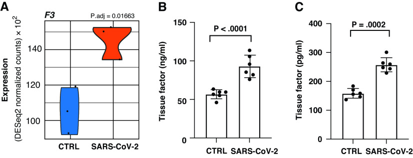
Figure 2.
(A) Violin plot depicting raw counts of reads mapping to key regulators of the extrinsic blood coagulation cascade in mock-infected and SARS-CoV-2–infected NHBEs as performed by Blanco-Melo and colleagues. Raw counts were normalized to library size in the DESeq2 software package. P.adj values for all differentially expressed genes were also calculated within DESeq2. Images were generated using GGPlot2 in the R studio environment. (B and C) ELISA measurements quantifying TF (tissue factor) in lysate and supernatant respectively. Samples were isolated at 24 hours after infection (multiplicity of infection 2) in NHBEs. Plotted values are the mean of two technical replicates ± SD. P values were determined using an unpaired two-tailed t test. CTRL = control.

