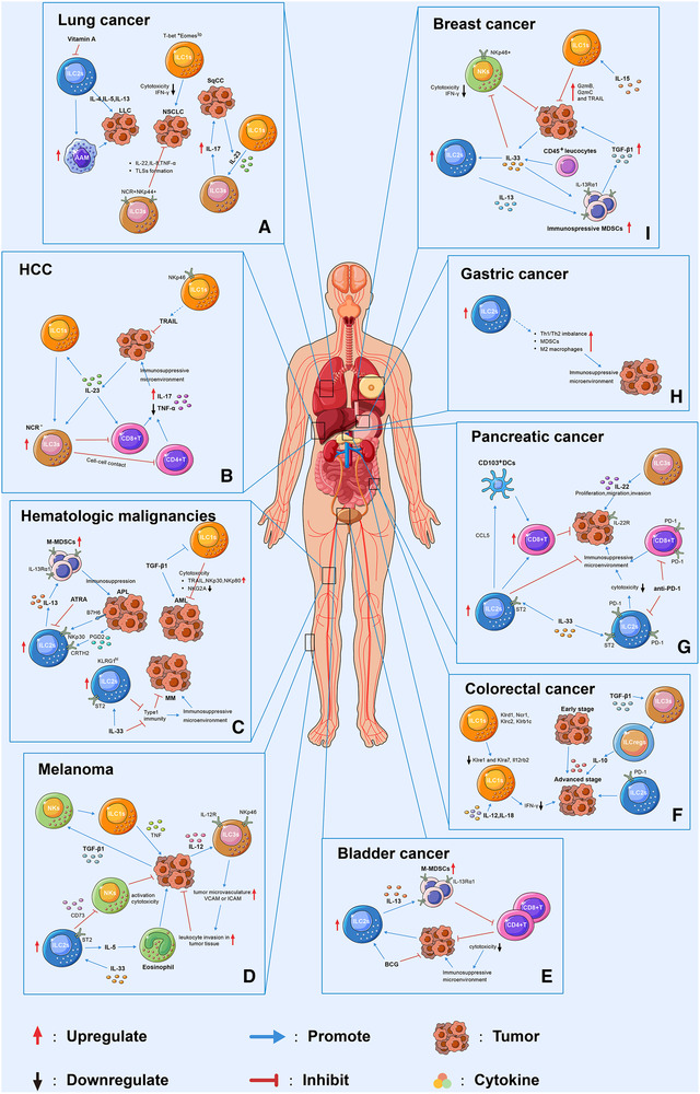
Figure 1.

Schematic representation of the dynamic behaviour of ILCs in different tumors and their interactions with the TME. Loss of function of ILC1s was associated with the tumor progression. For instance, the decreased cytotoxicity of ILC1s were reported in NSCLC (A), and in AML(C), melanoma (D), and breast cancer (I) in the presence of IL‐15 or TGF‐β. In the presence of IL‐23, ILC1s transdifferentiated into ILC3s which exhibited tumor progression function through producing IL‐17 in SqCC (A) and inhibiting CD4+ or CD8+ T cells in HCC (B). ILC2s were found to facilitate tumor progression through interacting with MDSCs by secreting IL‐13 in APL (C), bladder (E) and breast cancers (I), and with eosinophil through IL‐5 in the presence of IL‐33 in melanoma (D). In addition, ILC2s were shown to inhibited the cytotoxicity of NKs cell through CD73 in melanoma (D). In pancreatic cancer (G), ILC2s exhibit antitumor effect through attracting DCs by secreting CCL5, which lead to increase cytotoxic CD8+T cells in tumor microenvironment. Moreover, IL‐33 was capable of regulating ILC2s function in MM (C), pancreatic cancer (G), and breast cancer (I) through binding to its receptor ST2 on ILC2s. ILC2s might be associated with immunosuppressive environment in gastric cancer. For instance, increased frequency of ILC2s was correlated with Th1/Th2 imbalance, increase MDSCs and M2 macrophages in gastric cancer tumor tissue (H). The role of ILC3s is cancer type dependent. In NSCLC (A), ILC3s inhibit tumor progression through producing IL‐22, IL‐8, TNF‐α, and tertiary lymphoid structures (TLSs) formation at tumor site. In melanoma (D), IL‐2R+ILC3s recognize IL‐12 secreted by tumor cells and upregulate adhesion molecules in tumor vasculature, such as ICAM and VCAM, which lead to accumulated leukocyte in tumor tissue. In pancreatic cancer (G), ILC3s was associated with tumor progression, migration, and invasion. Single‐cell RNA sequencing revealed that ILC1s express activating receptors, such as Klrd1, Ncr1, Klrc2, and Klrb1c, at an early stage of CRC (F), and inhibitory receptors at a late stage such as Klre1 and Klra7. PD‐1high ILC2s was found in advanced stage of CRC and might be associated with tumor progression. Moreover, upon TGF‐β stimulation, ILC3s transdifferentiated into ILCregs in CRC, and deletion of IL‐10 in ILCregs suppressed tumor development.
