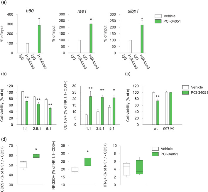
FIGURE 5.

HDAC8 inhibition increases NKG2D ligands in glioma cells boosting NK cell cytotoxicity. (a) ChIP experiments performed on CT2A cells treated or not with the HDAC8 inhibitor for 24 h using the indicated antibody (n = 3; *P < 0.05, Student's t‐test). (b) NK cells were incubated with CT2A cells pretreated with PCI‐34051 or vehicle for 48 h. degranulation of NK cells (having subtracted basal degranulation) in a 1:1, 2.5:1, and 5:1 E:T ratio with CT2A cells was assessed by FACS analysis of CD107a+ cells (right panel); cell viability in glioma cells is shown in the left panel (n = 6, *P < 0.05 **P < 0.01, one‐way ANOVA). Error bars show mean ± SEM. (c) NK cells, isolated from the spleen of wt or prf1 ko mice, were incubated with CT2A cells pretreated with PCI‐34051 or vehicle for 48 h. Glioma cell viability in a 2.5:1 E:T ratio is shown as mean ± S.E.M. (n = 6, **P < 0.01, one‐way ANOVA). (d) Percentage of CD69+, NKG2D+ and IFN‐γ+, cells in the CD3−/NK1.1+ cell population obtained from the brain of vehicle or PCI‐34051 treated mice (n = 4; *P < 0.05, Student's t‐test). For boxplots, the center line, boxes and whiskers represent the median, inner quartiles, and rest of the data distribution, respectively
