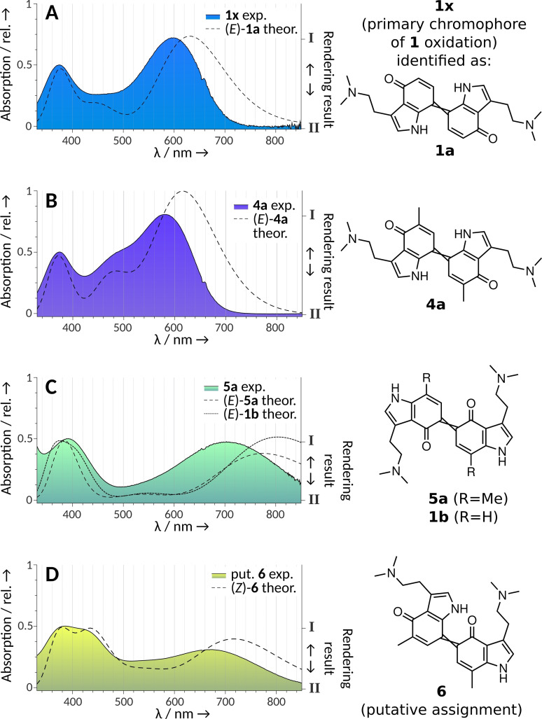
Figure 2.
Visual‐range diode array absorption spectra of quinoid dimers of 1 and of their 5‐ and 7‐methylated congeners 4 and 5, as well as comparison with their theoretical spectra calculated with time‐dependent density functional theory (dashed lines). The shading under the curves represents the actual substance colors under the chromatographic conditions (acetonitrile/H2O+0.1 % formic acid) depicted as color gradients between two rendered RGB values, obtained by two different computational methods (HDTV for I and Wα for II, see the Supporting Information). The right y‐axis refers to this gradient. A) The experimental spectrum represents the elusive putative dimer 1 x, which was later shown to be consistent with 1 a (=7,7’‐coupled unmethylated 1), i. e., the main dimeric blue compound of 1 oxidation. B) Dimer 4 a (=7,7’‐coupled, 5,5’‐dimethylated 4). C) Dimer 5 a (5,5’‐coupled, 7,7’‐dimethylated) and theoretical spectrum of 1 b (5,5’‐coupled unmethylated 1). D) Dimer 6 (5,7’‐coupled, 7,5’‐dimethylated 4 and 5).

