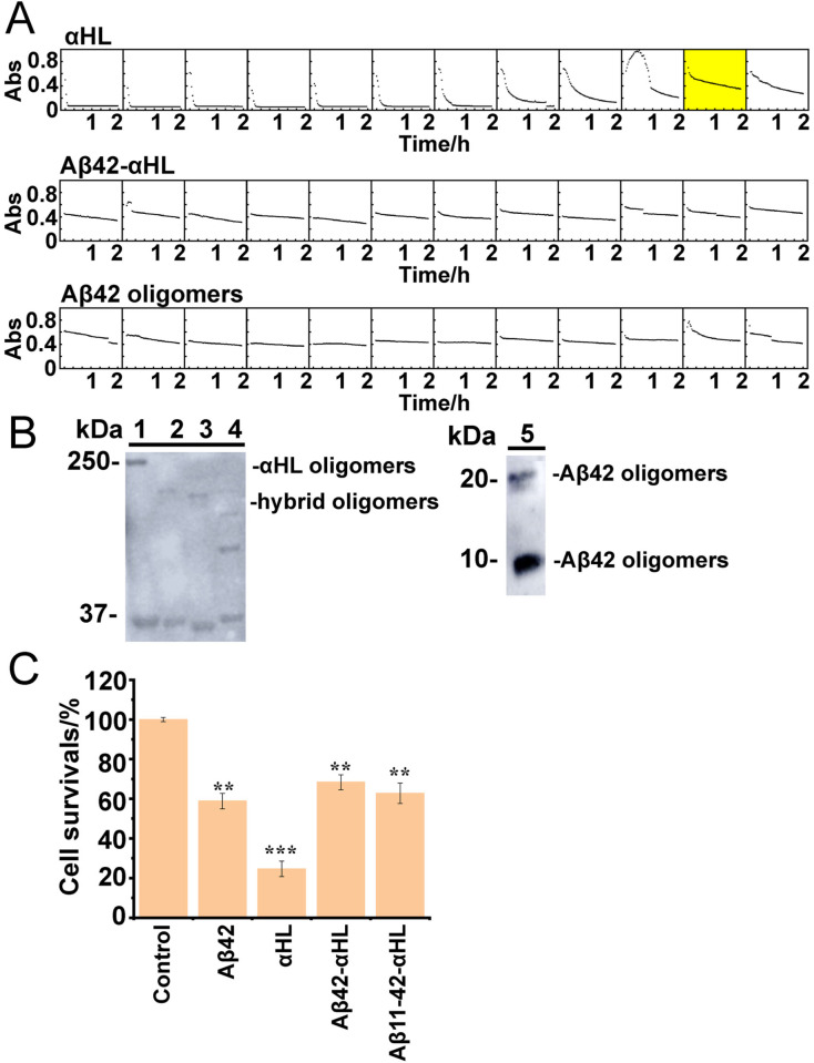
Figure 5.
A) Hemolysis by WT αHL oligomers, hybrid Aβ42‐αHL or WT Aβ42 oligomers oligomers. The HC50 of WT αHL (50 % cell lysis in 120 min at 37 °C; yellow box) is 24 ng mL−1, indicating specific hemolytic activity. The decrease of absorbance (Abs, y‐axis, from 0–1) in light scattering over time (x‐axis) was recorded in a microplate reader at 595 nm for 2 h at room temperature. B) Immunogenic similarity of WT αHL, hybrid Aβ42/11–42‐αHL and Aβ42 oligomers by western blot. The anti‐Aβ42 oligomer conformation‐dependent antibody A11 recognized αHL oligomers (lane 1); hybrid Aβ42‐αHL oligomers (lane 2); hybrid Aβ11‐42‐αHL oligomers (lane 3); hybrid hIAPP‐αHL oligomers (lane 4) and Aβ42 oligomers (lane 5). The SDS‐PAGE electrophoresis prior to blotting, was conducted at 120 V for 80 min. C) SH‐SY5Y cell viability using a luminescence assay (error bars determined by triplicate experiments). 5 μM WT αHL, Aβ42 and hybrid Aβ42‐αHL oligomers were treated with SH‐SY5Y cells with 6000 cells/well density after incubating for 48 h at 37 °C, 5 % CO2. The results are shown as percentages of control samples. All the Aβ42 oligomers were prepared in the presence of 0.38 mM DDM micelles. Data are represented as the mean S.E.M (standard error of the mean). Two‐tailed student's t‐test was applied for statistical significance. **P<0.01 (very significant) and ***P<0.001 (highly significant) were compared to the control.

