
Figure 3.
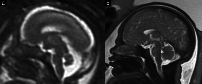
Comparison of the brain of an 18 + 5‐week (a) and a 36 + 1‐week (b) fetus on T2‐weighted magnetic resonance imaging. In the younger fetus, the areal contribution of the midbrain is significantly greater than that of the pons (41.7% vs 33.9%), whereas, in the older fetus, this relationship is reversed (32.3% vs 43.6%).