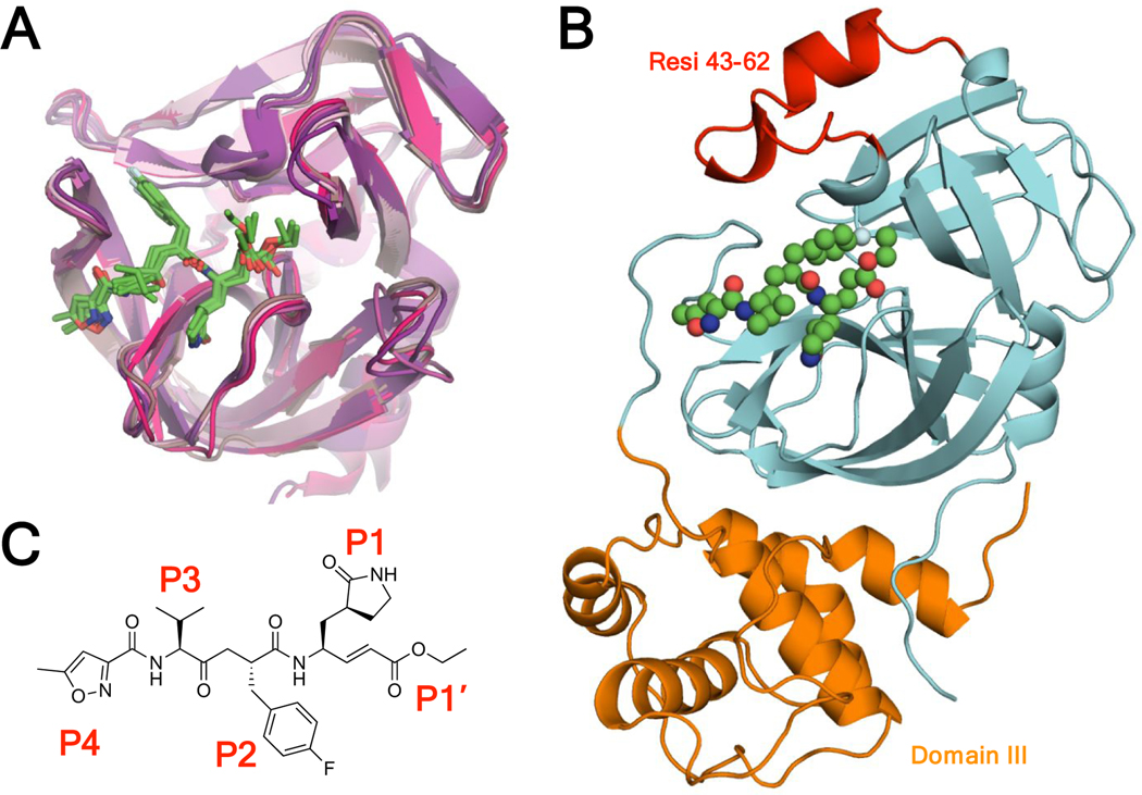
Figure 1.
A) Rupintrivir in complex with viral 3C proteases of rhinovirus A, rhinovirus C, coxsackievirus, enterovirus 71, enterovirus 93, and novel enterovirus 68. The crystal structures are superimposed in cartoon representation with rupintrivir shown as green sticks (PDB: 1CQQ, 3RUO, 3SJI, 3SJO, 6KU8, 7L8H). B) Novel SARS-CoV-2 Mpro-rupintrivir crystal structure in cartoon representation with rupintrivir shown as spheres, protease residues 43–62 colored red, the domain that is structurally homologous to 3C proteases colored cyan, and domain III colored orange (PDB: 7L8I). C) Chemical structure of rupintrivir.

