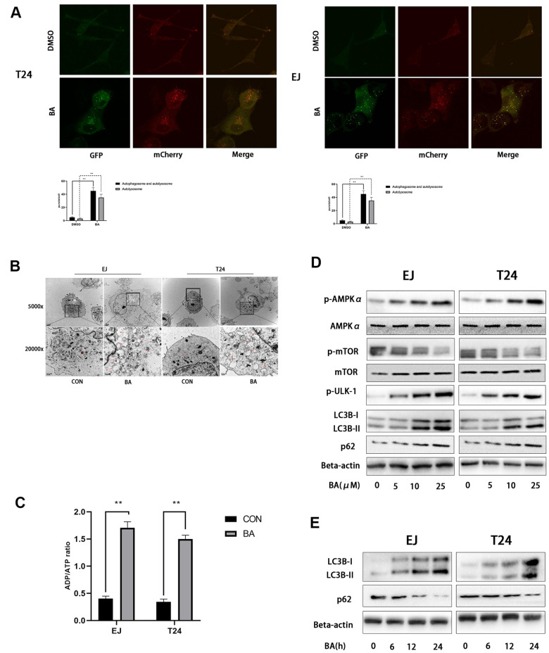
Figure 3.
BA induces autophagy in human bladder cancer cells. (A) Confocal fluorescence microscopy of EJ and T24 cells transfected with mCherry-GFP-LC3B lentivirus and exposed to 25 μM BA for 24 h. (B) Transmission electron microscopy (magnification: 5000× and 20,000×) images of EJ and T24 cells exposed to 25 μM BA for 24 h. Arrows indicate autophagic vesicles. (C) Luminescent determination of ADP/ATP ratio in EJ and T24 cells exposed to DMSO (control) or BA for 24 h. (D, E) Western blot analysis of changes in AMPK-mTOR-ULK1 phosphorylation status and p62 and LC3B-II expression upon treatment with the specified BA doses. *p<0.05, **p<0.01.

