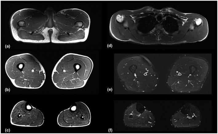
FIGURE 5.

(a–f). Axial T1‐SE (a,b,c,d) and T2‐STIR sequences (e and f) of the pelvic girdle (a), shoulder girdle (d), thigh (b and e), leg (c and f), bilaterally. Muscle trophism and signal are normal at the level of the thigh (b) and shoulder girdle (d), whereas slight hyperintensity can be detected at the level of glutei maximi (Mercuri score 1) (a) and of the muscles of the posterior compartment of the legs (c). No muscle edema could be detected in the inferior limbs (e and f)
