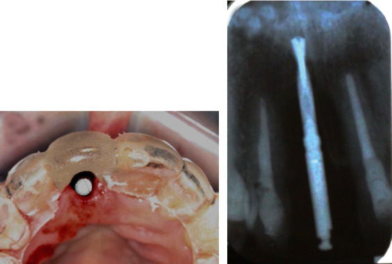
Figure 3.

(a) Initial implant osteotomy was made at the cingulum region using a precision drill under the guidance of a surgical stent. (b) A periapical radiograph was taken with a 2 mm twist drill to verify the depth and angulation for implant placement.
