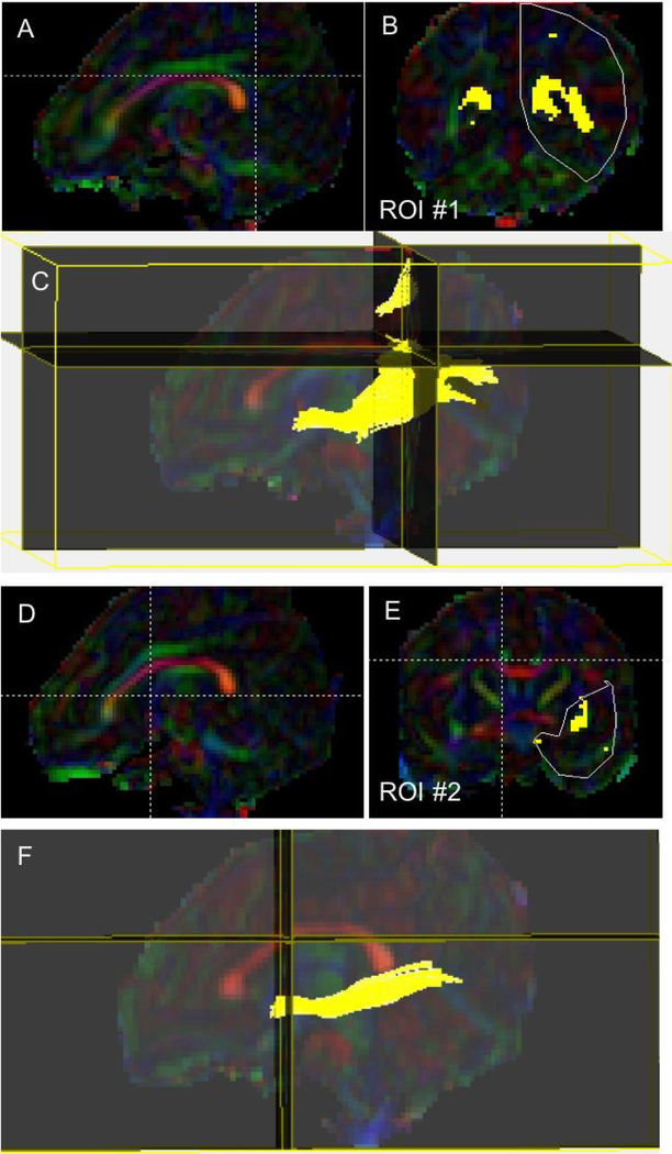
Figure 1.
Inferior longitudinal fasciculus (ILF) segmentation: (A) The sagittal color map image that was used to identify the location of the first region of interest (ROI) at the level of the posterior edge of the intensely green cingulum region in a representative ELBW infant; (B) Coronal image showing the first polygonal ROI covering the entire left hemisphere; (C) 3D sagittal view of the ILF fiber bundle after placement of the first ROI; (D) Sagittal image used to identify the second ROI at the level of the anterior third of the genu of the corpus callosum; (E) Coronal image showing the second polygonal ROI; (F) 3D trajectory of the final IFL fibers after placement of the second ROI and fiber exclusion protocol.

