
Figure 6: Demonstration of S1P1 (A, B) or SR-BI (C, D) in the endothelium of aortas from Apoe haploinsufficient mice without (CTRL: A, C) or with overexpression of S1P1 (S1P1-iECKI; B, D).
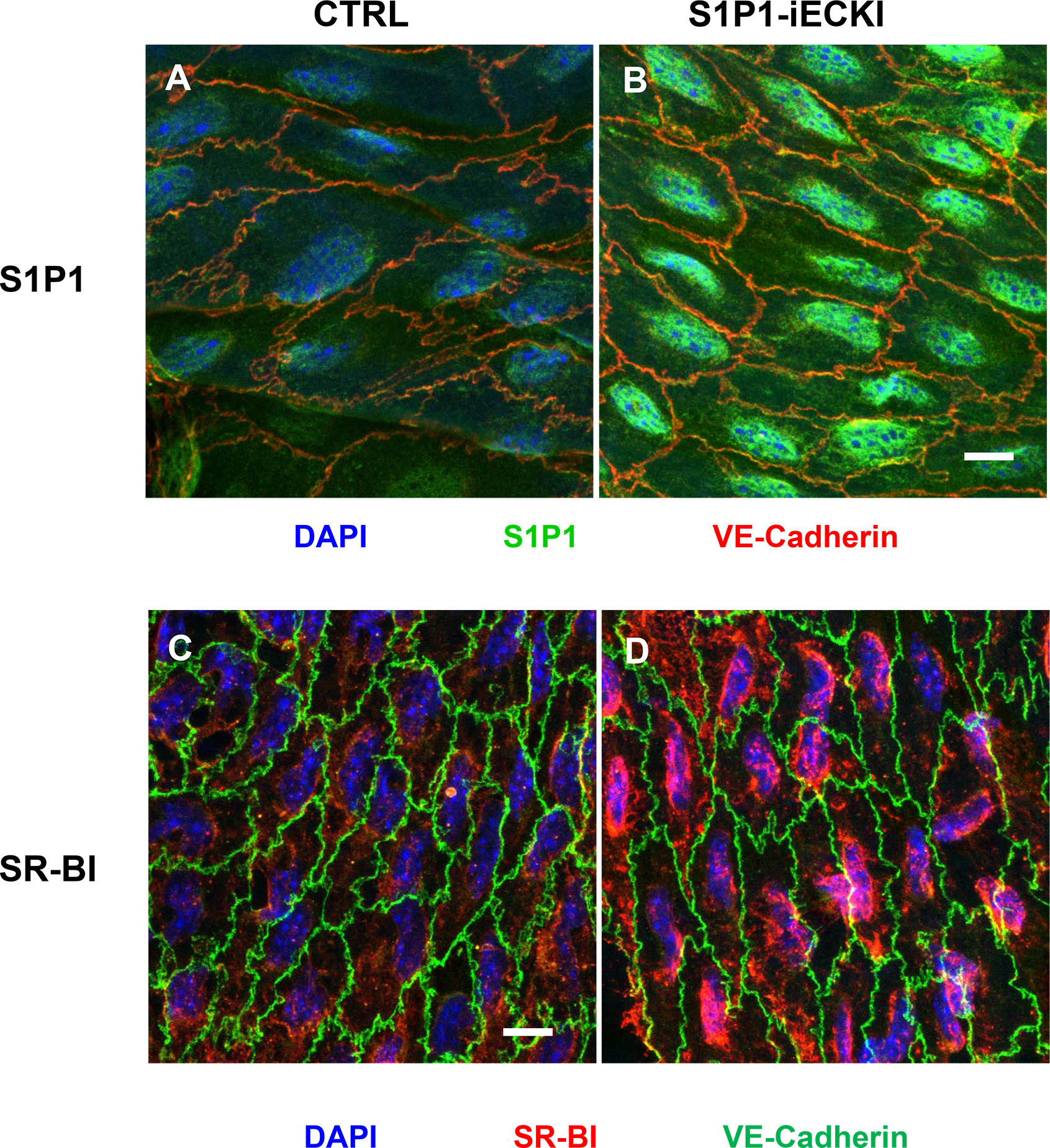
Figure shows en-face prepared aortic immunostainings. Aortas were quickly cleaned of adventitial tissue, opened longitudinally and incubated with primary and secondary antibodies conjugated with green or red fluorescent dyes, as indicated. Nuclei were counterstained with DAPI. Images were captured by confocal microscope and z-axis projections of 14 scanned planes are shown. Scale bar = 10μm. Original micrographs are shown as supplemental figures IV and V.