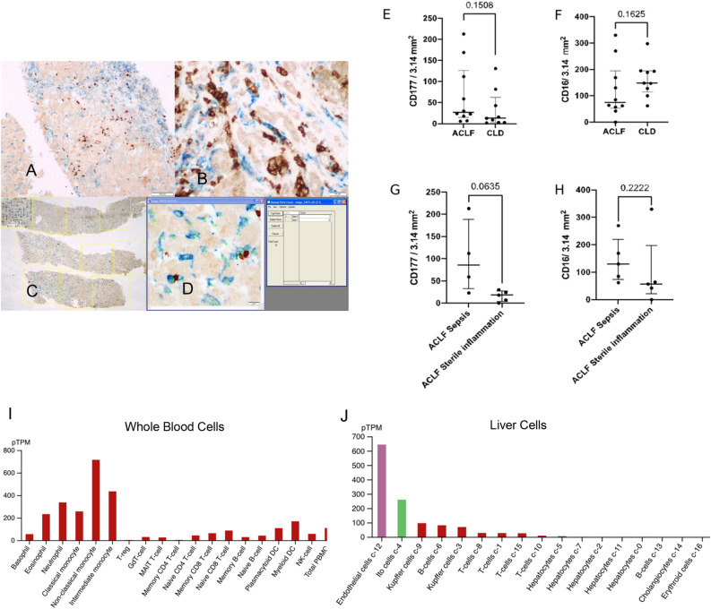
Figure 5.
Immunohistochemistry of post-mortem biopsies obtained from ACLF and CLD-AD patients. (A, B) The dual-colour stained slides were photographed by using a BX43 Olympus microscope. The CD177 stained neutrophils were represented by brown stain (black arrows) and the CD16 stained neutrophils, monocytes, macrophages, and Kupffer cells were represented by blue stain (blue arrows) (A × 40, B × 400). (C) Method for observation of dual-colour stained slide under 2 × objective of the microscope and selection of non-overlapping FOVs followed by counting at × 40; (D) Method of using the manual tagging and counting tool of the Image Proplus 6.1 software [D × 40] (E) CD177+ Neutrophil count/ 3.14 mm2 in ACLF and CLD-AD patients; (F) CD16+ leukocytes in ACLF and CLD-AD patients; (G) CD177+ Neutrophil count/ 3.14 mm2 in ACLF patients with or without sepsis; (H) CD16+ leukocytes in ACLF and CLD-AD patients. Sample size for each group- ACLF (n = 10); CLD-AD (n = 9); ACLF sepsis (n = 4); ACLF sterile inflammation (n = 5). For 1 ACLF sample, categorization as sepsis/ sterile inflammation was not available). Non-parametric Mann Whitney Test was performed to determine p values (indicated on the top of the graphs). Graphs are plotted as Median with IQR. (I) Cell-type specific expression of CD177 binding partner PECAM1 in whole blood cells. (J) Cell-type specific expression of PECAM1 in liver cells. (I, J) Data have been taken from Human Protein Atlas.

