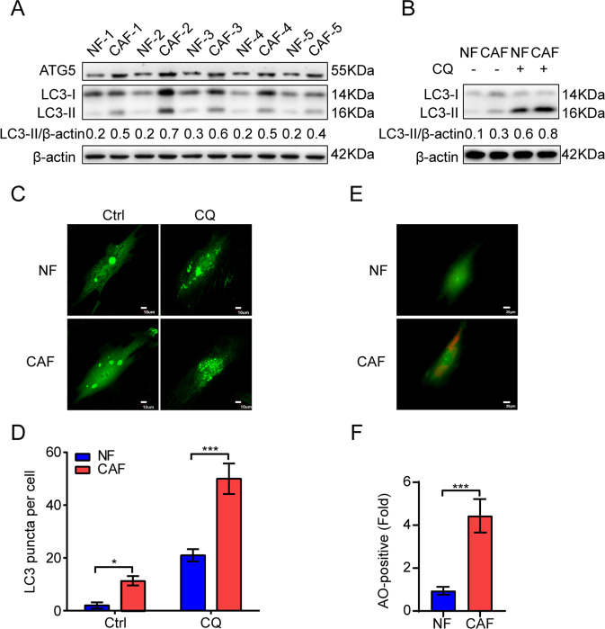
Fig. 1. CAFs of lung cancer possess a high basal level of autophagy.
A The expressions of ATG5 and LC3 in paired CAFs and NFs were detected by western blotting. (n = 5). B CAFs and NFs were treated with CQ (60 µM) for 2 h, the expression of LC3 was detected by western blotting. C CAFs and NFs were infected with Ad-GFP-LC3. After 24 h, cells were treated with CQ (60 µM) for 2 h. LC3 puncta patterns were observed under a confocal microscope. Scale bar, 10 μm. D Quantitative analysis of GFP-LC3 puncta. E CAFs and NFs were incubated with AO (1 μg/mL) for 15 min. The formation of acidic vesicles organelles (AVOs) was observed under a confocal microscope (×400 magnification). Scale bar, 20 μm. F Quantitative analysis of formation of AVOs. Data represent the mean ± SD from three independent experiments. Columns, mean; bars, SD. *p < 0.05, ***p < 0.001. Ctrl control.

