Figure 1.
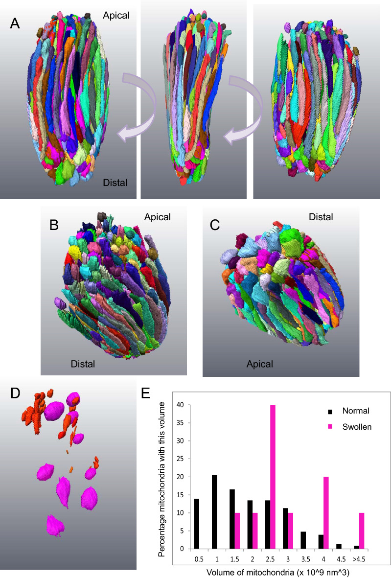
3D rendering of complete segmentation of every mitochondrion in a small, healthy-looking cone. (A) Three panels represent 90 degree rotations of the ellipsoid of a cone cell showing all the mitochondria. The mitochondria show a range of profiles, though most are long and thin. (B) Mitochondria at the apical end of the ellipsoid terminate with narrow tips. There were also numerous mitochondrial fragments. (C) Those at the distal end were swollen at their tip. (D) Mitochondrial fragments (red) are mostly localised to the apical domain of the ellipsoid. Spheroidal, swollen mitochondria (magenta) are found throughout. (E) Histogram showing the range of mitochondrial volumes in this ellipsoid. The spheroidal mitochondria (magenta) have a greater average volume.

