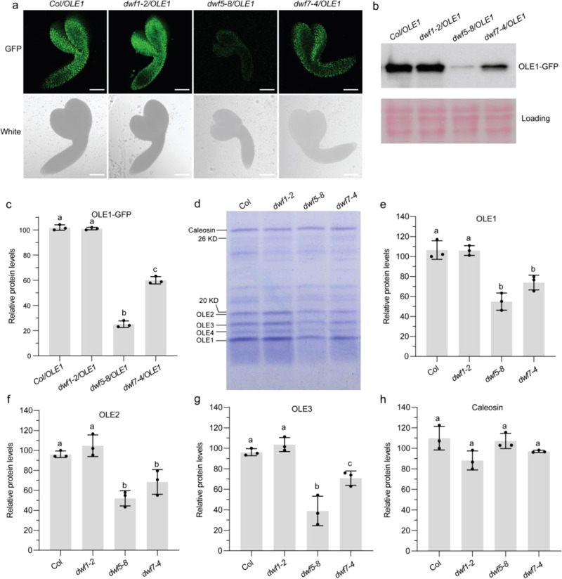
Fig. 7. Seed oleosin protein abundance is decreased in dwf5 and dwf7 mutants.
a Representative images of LDs (OLE1-GFP, green) in embryos at the bent cotyledon stage. The experiment was repeated three times with similar results. Bar = 100 μm. b Western blotting with anti-GFP antibody showing OLE1-GFP protein levels in embryos of Col/OLE1, dwf1-2/OLE1, dwf5-8/OLE1, and dwf7-4/OLE1 at the bent cotyledon stage. Equal loading of proteins are shown by Ponceau S staining. c Relative OLE1-GFP protein levels quantified using Image J software. d–h, Oleosin and caleosin protein levels in seed LDs. LDs were purified from dry seeds. Proteins of LDs equivalent to 15 μg of TAGs were subjected to SDS-PAGE with Coomassie blue staining (d). Relative oleosin and caleosin protein levels were quantified using ImageQuant TL software (e–h). In c, e, f, g, and h, data are mean ± SD. of three biological replicates. Different letters indicate significant differences at P < 0.05, as determined by one-way ANOVA with Tukey’s multiple comparisons test.

