图 1.
A typical case
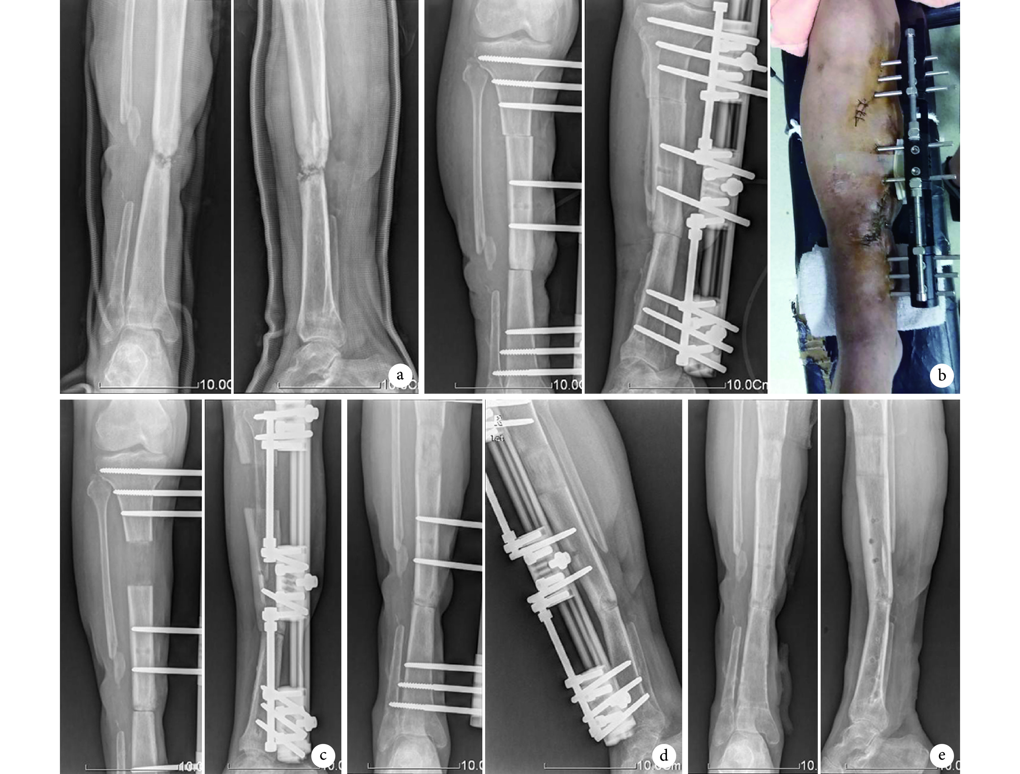
典型病例
a. 术前正侧位 X 线片示胫骨中段慢性骨髓炎并病理性骨折;b. 病灶骨节段切除短缩术后正侧位 X 线片及患肢外观;c. 术后肢体延长 2 个月正侧位 X 线片;d. 术后 6 个月正侧位 X 线片示对合骨端骨痂及牵张骨痂生长;e. 术后 10 个月去除外固定后正侧位 X 线片示延长端及对合端均达骨性愈合
a. Anteroposterior and lateral X-ray films before operation, showing chronic osteomyelitis of tibia and pathological fracture; b. Anteroposterior and lateral X-ray films and appearance after excision of lesion bone and shortening; c. Anteroposterior and lateral X-ray films after limb lengthening for 2 months; d. Anteroposterior and lateral X-ray films at 6 months after operation, showing callus growth in the opposite end of bone; e. Anteroposterior and lateral X-ray films after removal of external fixator at 10 months after operation, showing bone healing of extended end and opposite end of bone