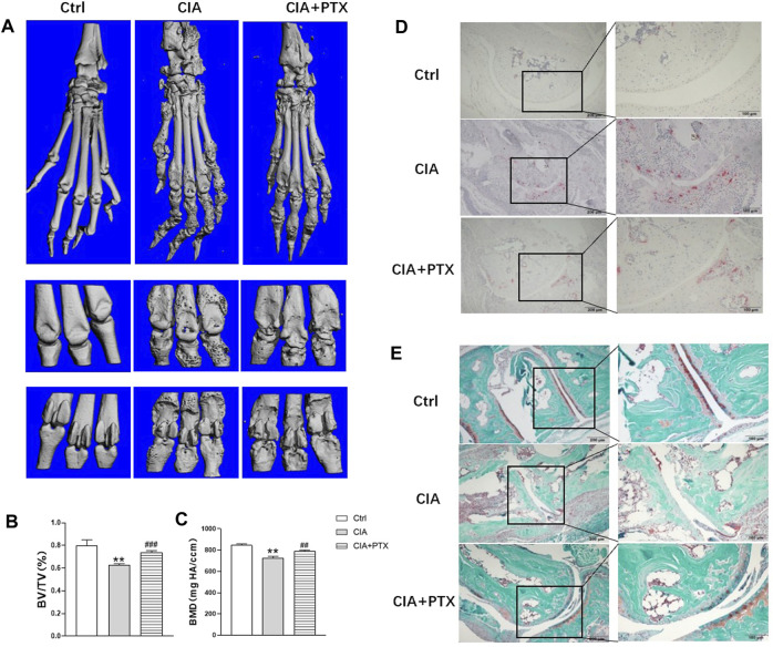
FIGURE 6.
PTX protects against bone destruction in CIA mice. (A) Micro-CT scanning images of paws and joints. (B–C) The bone volume/tissue volume (BV/TV) and bone mineral density (BMD) were analyzed. (D) TRAP staining was used to evaluate osteoclasts. (E) Safranine-O/fast green staining was employed to assess cartilage damage. Data are expressed as mean ± SD (n = 5). * p < 0.05, ** p < 0.01 vs Ctrl group. # p < 0.05, ## p < 0.01 vs CIA group.

