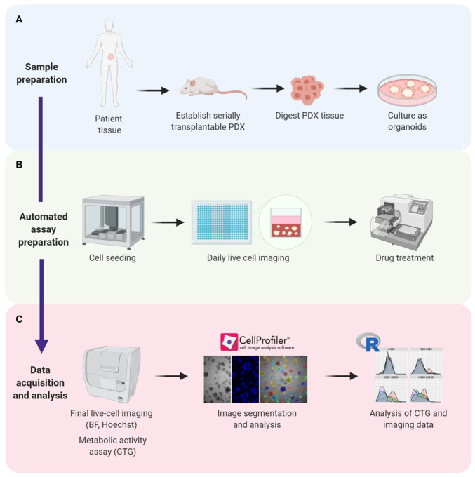
Figure 1.
Establishing a high-throughput assay for automated seeding, treatment, and analysis of prostate cancer organoids. (A) Prostate cancer tissue is acquired from patients and used to establish serially transplantable patient-derived xenografts (PDXs). PDX tissue is then digested and cultured in Matrigel as organoids. (B) Organoids are robotically seeded in 384-well plates and monitored at different intervals using live-cell brightfield microscopy. Drug treatment started at 8 d in culture and concluded at day 21. (C) After treatment, live-cell imaging followed by endpoint CTG measurements were performed. Microscopy images were segmented and quantified using CellProfiler software. CTG and imaging data were analyzed using R software. BF = brightfield microscopy; CTG = CellTiter-Glo; PDX = patient-derived xenograft.

