FIGURE 1.
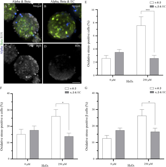
The addition of endothelial cells in the pseudoislet reduces the oxidative stress in α and β cells (A, B) Fluorescence imaging reveals ROS-positive cells in samples that were exposed to 250 µM H2O2, showing α cells (blue), β cells (green) and ROS (grey) (C, D) Oxidative stress varies in pseudoislets with and without endothelial cells. A lower percentage of oxidative stress-positive cells are found in the pseudoislets including endothelial cells. Scale bars: 50 µm (E) After 5 days in culture, the endothelial cells decrease oxidative stress (***p < 0.001) (F) The presence of endothelial cells in the pseudoislet did not affect the percentage of oxidative stress-positive α cells in the basal media, however when induced by H2O2, a significant decrease of oxidative stress-positive α cells was seen (*p < 0.04) (G) The inclusion of endothelial cells in the pseudoislet reduced the percentage of oxidative stress-positive β cells upon induction by H2O2 (*p < 0.04). Results are expressed as mean ± SEM, and each data set includes twelve pseudoislets (n:12), and the experiment was repeated three times (N:3).

