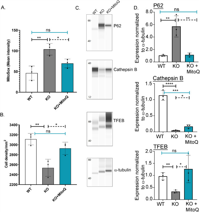
Figure 7.
MitoQ in vivo reduces mitoROS, decreases cell loss, and improves lysosome protein expression in CHED mice. Intra-peritoneal injections of MitoQ (68 µg) on alternate days starting at 8 weeks of age for 4 weeks. (A) MitoSox fluorescence intensity in corneal endothelium of WT, KO, KO + MitoQ animals, n = 3. Two females and one male mice for each group was used for this assessment. (B) Corneal endothelial density, n = 3, two female mice and one male mouse from each group. (C) Wes immunoassay of P62, Cathepsin B, and TFEB from Slc4a11 WT and KO animals, two female mice of each group, repeated three times (n = 2 animals per group for each of the 3 trials). (D) Quantification of panel C data, mean ± SD, *P < 0.05, **P < 0.01, ***P < 0.001, ****P < 0.0001 (1-way ANOVA with Tukey's multiple comparisons test).

