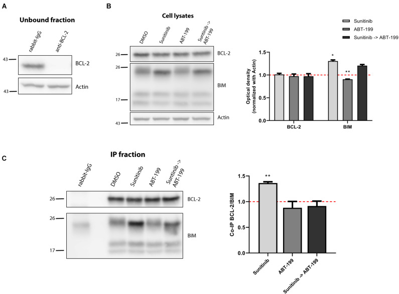
FIGURE 6.
BCL-2 binds to BIM after sunitinib treatment promoting synergy with ABT-199. (A) Western blot analysis of the unbound fraction after BCL-2 immunoprecipitation. (B) Immunodetection of BCL-2 and BIM initial expression in cell lysates after 16 h of incubation with sunitinib 100 nM, and 4 h incubation with ABT-199 10 nM. Quantification of optical density for each protein was normalized to actin and fold-change was calculated comparing to protein expression in the control condition. (C) Western blot of the immunoprecipitated fraction to study the interaction between BCL-2 and BIM after sunitinib 100 nM treatment, and 4 h with ABT-199 10 nM. To quantify this binding, BIM optical density was normalized to BCL-2 optical density and fold-change was calculated comparing to protein expression in the control condition. All results are expressed as the mean ± SEM of at least three biologically independent replicates. Statistical significance was calculated using Student’s t-test compared to control condition and considering *p < 0.05 and **p < 0.01.

